42 brain cross section diagram
Which shape is a possible representation of the cross section? - brainly.com The diagram shows a sphere with center O and a radius of 4 Inches that has been cut by a plane. The cross-section formed by the plane is shaded. Which shape is a possible representation of the cross section? 1 See answer Advertisement avacodosamkapoor is waiting for your help. Add your answer and earn points. facundo3141592 Answer: A circle. Body Planes and Sections: Anatomical Position, Directional Term Definitions ... Anatomical position, body planes, anatomy sections. Directional term descriptions, definitions, example labeled diagrams of sagittal, coronal, transverse, oblique, and longitudinal axis. Quiz yourself on how each plane divides the body into front (anterior) and back (posterior) portions, right and l
Midbrain | Radiology Reference Article | Radiopaedia.org The midbrain, or mesencephalon (plural: mesencephala or mesencephalons), is the most rostral part of the brainstem and sits above the pons and is adjoined rostrally to the thalamus.During development, the midbrain forms from the middle of three vesicles that arise from the neural tube.. When viewed in cross-section, the midbrain can be divided into three portions:
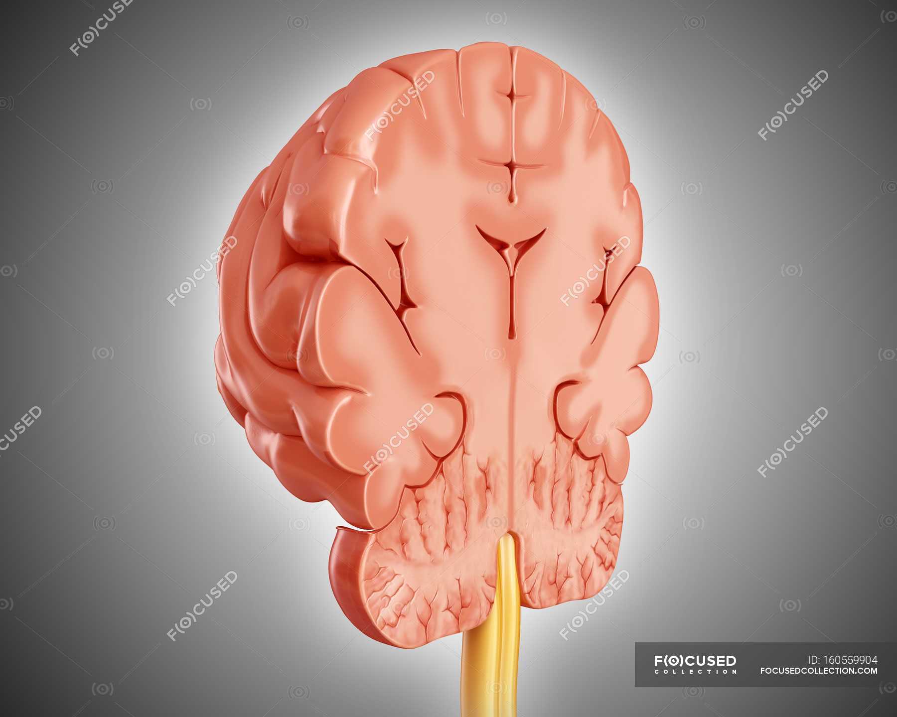
Brain cross section diagram
Draw a neat well labelled diagram of a human eye. Human eye, in humans, specialized sense organ capable of receiving visual images, which are then carried to the brain. cross section of the human eye. A horizontal cross section of the human eye, showing the major parts of the eye, including the protective covering of the cornea over the front of the eye.Made of many working parts, the human ... Semester 2 Neuro , Quiz 7 - CNS Blood Supply - ProProfs Semester 2 Neuro , Quiz 7 - CNS Blood Supply. . 1. A 55-year old male was admitted to the hospital after suffering from a stroke. He later succumbed. The diagram below represents a cross section from his lower pons obtained at autopsy. Sections Of The Brain - what part of the brain coordinates movement and controls ... Sections Of The Brain - 17 images - physiology of the brain and it s various functions, cta of the brain with bone removal via de technique, cta of the brain with bone removal via de technique, brain frontal section 2 human pathology,
Brain cross section diagram. Brain: Ultimate Guide to the Brain for AP® Psychology The Lobes of The Brain Diagram showing the lobes of the brain. Image Source: Wikimedia Commons. The parietal lobe is located at the top of the brain, between the frontal and occipital lobe. It consists of the somatosensory cortex and is responsible for integrating sensory information from different parts of the body, especially visual ... Brain Wiring Diagram - easywiring Given below is a diagram of brain its functions detailing the four lobes and their associated structures. By the age of 80 the total length of wiring in the male brain has shrunk to 97 200 km and the total axon length in the aged female brain has decreased to 82 00 km. It is separated from the parietal lobe by the central sulcus. Optic Nerve: Anatomy, Location, and Function Anatomy. The optic nerve is mainly made up of the axons (nerve fibers) of the retinal ganglion cells from the retina. The optic disc or nerve head is the point where the axons from the retinal ganglion cells leave the eye. The nerve head appears as a white circular structure in the back of the eye. There are no photoreceptors on this structure. Spinal Cord Cross Section Explained (with Videos) - New Health Advisor Spinal Cord Cross Section Looking at a cross section of the spinal cord, you would see gray matter shaped like a butterfly surrounded by white matter. The gray matter is the core and ends up to be four projections that are known as horns. At the back are two dorsal horns and away from the back are two ventral horns.
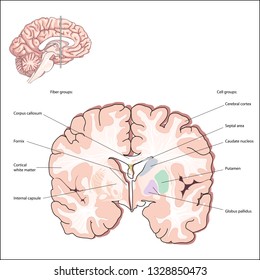
Cross Sectioning - cross section views, typical cross section download scientific ... Cross Sectioning. Here are a number of highest rated Cross Sectioning pictures on internet. We identified it from well-behaved source. Its submitted by government in the best field. We recognize this nice of Cross Sectioning graphic could possibly be the most trending topic afterward we share it in google improvement or facebook. › self-awareness › personalityPersonality Theories and Types - BusinessBalls.com Jun 09, 2017 · Typically this will equate to the Jungian 'superior function' and the Myers Briggs® 'dominant function' as described in this section. Benziger's books ('The Art of Using Your Whole Brain', and in revised form 'Thriving in Mind') contain an excellent and simple personality assessment to illustrate this point. Cross-sectional anatomy of the brain - e-Anatomy - IMAIOS Cross-sectional anatomy of the brain - e-Anatomy Brain - MRI (Axial) Sagittal Coronal 3D 1/24 Revert to the old version of the viewer e-Anatomy Authors Antoine Micheau , Denis Hoa Published on Friday 15 April 2022 Section Brain DOI ISSN 2534-5079 Anatomical parts Angular gyrus Anterior cerebral artery › human-body-maps › corpus-caverCorpus Cavernosum Penis Anatomy, Function & Diagram | Body Maps Mar 31, 2015 · The penis is composed of three cylinders encased in a sheath called the bucks fascia. These three cylinders are the corpus spongiosum and two corpora cavernosa known as the corpus cavernosum of penis.
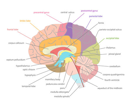
Anatomical diagrams of the brain - e-Anatomy - IMAIOS Neuroanatomy : Brodmann areas, Broca area, Wernicke area. Three anatomical sections of the brain (axial, coronal and sagittal) close this chapter on the brain. Brain , Coronal section : Brain , Anatomy diagram. Numerous illustrations are available on the cerebellum, representation of cerebellar lobes, fissures, sulci and the vermis. Midsagittal Brain Diagram - brain anatomy nursing care plan nursing pedia, brain ... Midsagittal Brain Diagram. Here are a number of highest rated Midsagittal Brain Diagram pictures upon internet. We identified it from well-behaved source. Its submitted by supervision in the best field. We believe this nice of Midsagittal Brain Diagram graphic could possibly be the most trending ... The diagram below shows a cross-section through an alveolus and a capillary. Why ... The diagram below shows a cross-section through an alveolus and a capillary... The diagram below shows a cross-section through an alveolus and a capillary. Why does oxygen move from P to Q? Answers: 2 Get Other questions on the subject: Biology. Biology, 21.06.2019 17:00, antcobra ... 📈 The diagram shows a wooden prism of height 5cm. The cross section of the prism ... The cross section of the prism is a sector of a - Brainly.com nathreesya 07/23/2021 Mathematics College answered The diagram shows a wooden prism of height 5cm. The cross section of the prism is a sector of a circle with sector angle 25º. The radius of the sector is 15 cm. Calculate the total surface area of the prism. Advertisement
Human brain cross-section — anatomy reference, 3d - Stock ...
Labelled imaging anatomy cases | Radiology Reference Article - Radiopaedia URL of Article. This article lists a series of labelled imaging anatomy cases by body region and modality. On this page: Article: Brain. Head and neck. Spine. Chest. Abdomen and pelvis.

Human brain cross-section, illustration - Stock Image - F016 ...
Cross sectional anatomy - Kenhub Cross section through the thalamus: Diagram Orienting yourself within such a cross section is easy. The star of the show (brain) is easily recognizable because it appears highly convoluted, full of ridges (gyri) and indentations (sulci). The paired thalami appear as two circular masses in the midline, forming the walls of the third ventricle.

Cross section of brain with arteries." Canvas Print by ...
Earthworm Dissection Internal View, Brain Labeled Brain. Cross-Section of Earthworm Segment. Cut a thin cross-section through your worm's intestinal area and view it with a dissecting scope. Also, view the prepared slide of an earthworm cross section (Carolina #Z1250), and examine the large model worm. Cross Section, Whole Worm Labeled Worm Cross Section
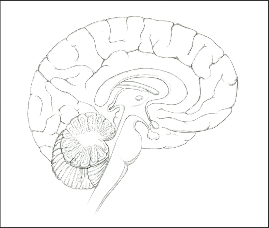
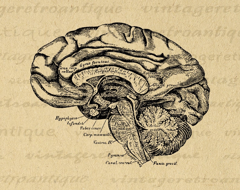
Sagittal Cross Section of the Brain - Amanda Barnaby
Body Cavities Labeled: Organs, Membranes, Definitions, Diagram, and Lateral View ... The spinal meninges are similar to the brain. We can use the circular cross-section below as a reference. The cross-section illustrates as if we are looking down at the spinal cord, and it shows the layers of the spinal cavity discussed above. The spinal cord shown in red is in the center of the spinal cavity.

Brain Cross section Diagram | Quizlet
Hippocampus - Wikipedia Image 2: Cross-section of cerebral hemisphere showing structure and location of hippocampus Image 3: Coronal section of the brain of a macaque monkey, showing hippocampus (circled) The hippocampus can be seen as a ridge of gray matter tissue , elevating from the floor of each lateral ventricle in the region of the inferior or temporal horn.

Human Brain. Cross Vector & Photo (Free Trial) | Bigstock
Human Brain Diagram Blank - brain anatomy stock photos images pictures ... Human Brain Diagram Blank - 17 images - brain outline clip art at vector clip art, the 25 best brain diagram ideas on pinterest diagram of, human skeleton blank clip art at vector clip, exploring the human brain how you will be graded assessment,

Human brain anatomy diagram cross section with all lobes and ...
Labeled Diagram Of Endocrine System - Hormones Booklet Docx Label The Major ... The key endocrine glands and organs are listed below: Thyroid glands, testes, pancreas, ovary, brain, adrenal glands. Medical diagram with closeup gland cross section. In this video, i will draw the well labelled diagram of human endocrine glands of both male and female.this video is about to draw the . The major glands of the .
Human brain cross-section — nervous system, central nervous ...
Brainstem: Definition, anatomy, parts, function - Kenhub Cross section of medulla at the level of the vagus nerve (overview diagram) The nuclei at the dorsolateral part of the medulla have (arranged from medial to lateral) the posterior (dorsal) vagal, solitary, medial vestibular, and cuneate nuclei. The cuneate nucleus is situated within the inferior cerebellar peduncle.
Cross Section Of Brain Illustration 60816752 - Megapixl
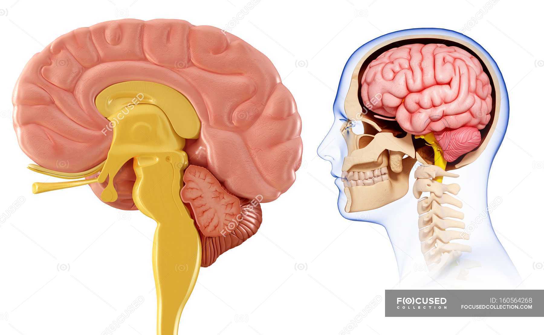
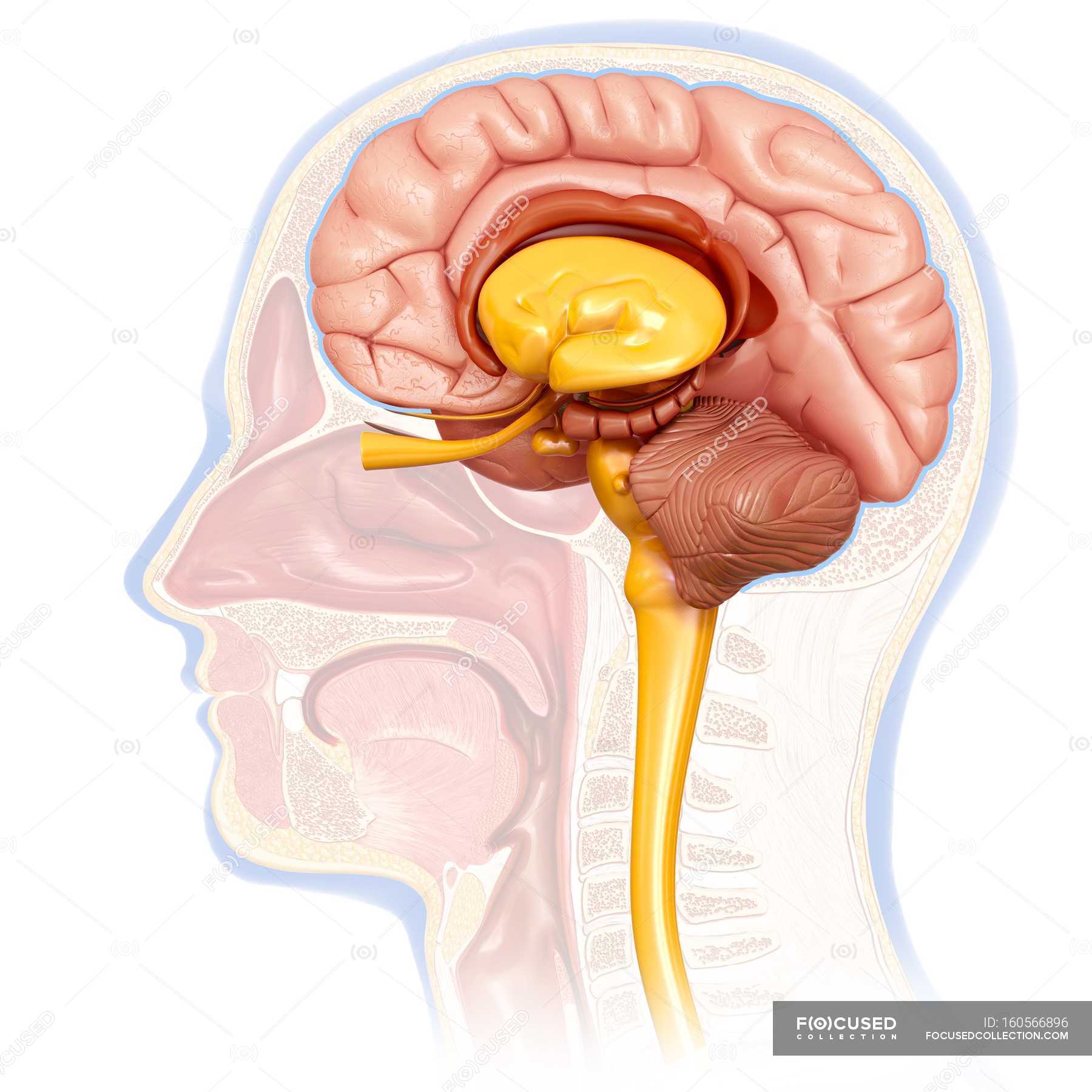
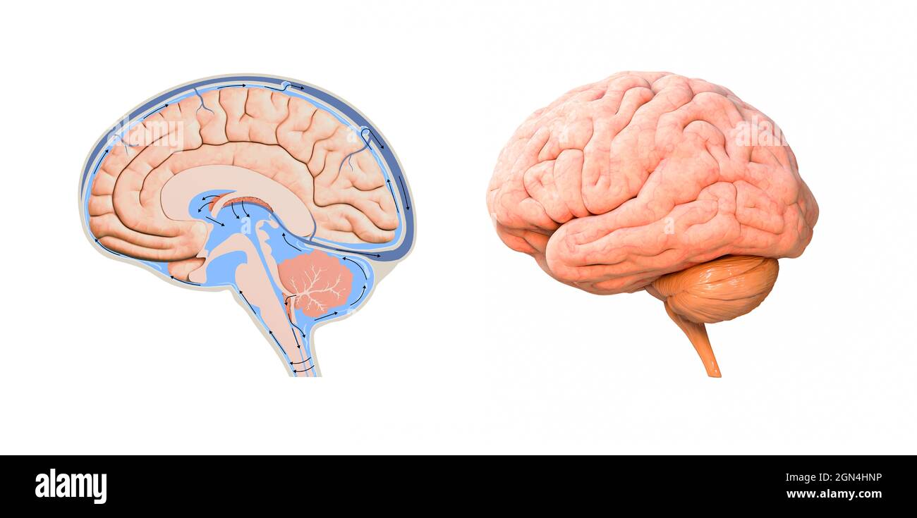
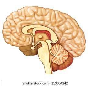
Chapter 16. Brain | The Big Picture: Gross Anatomy | AccessMedicine | McGraw Hill ... Medial view of the sagittal section of the brain. Cerebrum The cerebrum is the organ of thought and serves as the control site of the nervous system, enabling us to possess the qualities associated with consciousness such as perception, communication, understanding, and memory ( Figure 16-1A ).

Brain And Blood Vessels Of The Brain, Beautiful Colorful ...
Sections Of The Brain - what part of the brain coordinates movement and controls ... Sections Of The Brain - 17 images - physiology of the brain and it s various functions, cta of the brain with bone removal via de technique, cta of the brain with bone removal via de technique, brain frontal section 2 human pathology,

Cross-sectional view of the brain - Stock Illustration ...
Semester 2 Neuro , Quiz 7 - CNS Blood Supply - ProProfs Semester 2 Neuro , Quiz 7 - CNS Blood Supply. . 1. A 55-year old male was admitted to the hospital after suffering from a stroke. He later succumbed. The diagram below represents a cross section from his lower pons obtained at autopsy.
Human brain cross-section — internal organ, anatomy - Stock ...
Draw a neat well labelled diagram of a human eye. Human eye, in humans, specialized sense organ capable of receiving visual images, which are then carried to the brain. cross section of the human eye. A horizontal cross section of the human eye, showing the major parts of the eye, including the protective covering of the cornea over the front of the eye.Made of many working parts, the human ...
Cross section of brain Images, Stock Photos & Vectors ...
Brain Anatomy Images – Browse 147,965 Stock Photos, Vectors ...

Cross section brain Images, Stock Photos & Vectors | Shutterstock
Printable Image Brain Diagram Cross Section Digital Medical ...
![Brain cross section - Stock Illustration [65838488] - PIXTA](https://en.pimg.jp/065/838/488/1/65838488.jpg)
Brain cross section - Stock Illustration [65838488] - PIXTA
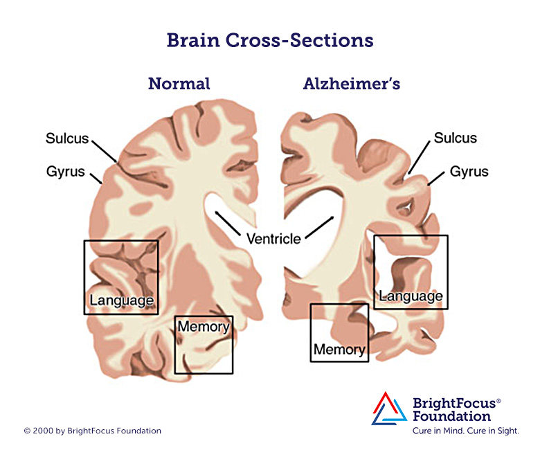
Brain with Alzheimer's Disease | BrightFocus Foundation
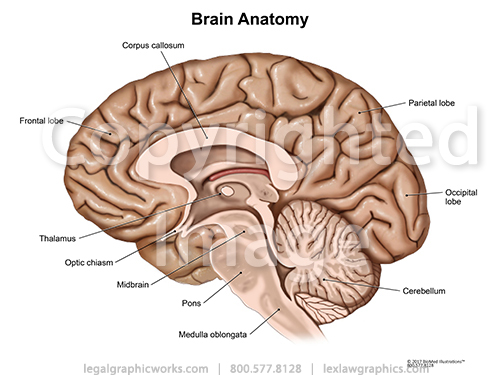
Cross Sectional Brain Anatomy - Legal Graphicworks

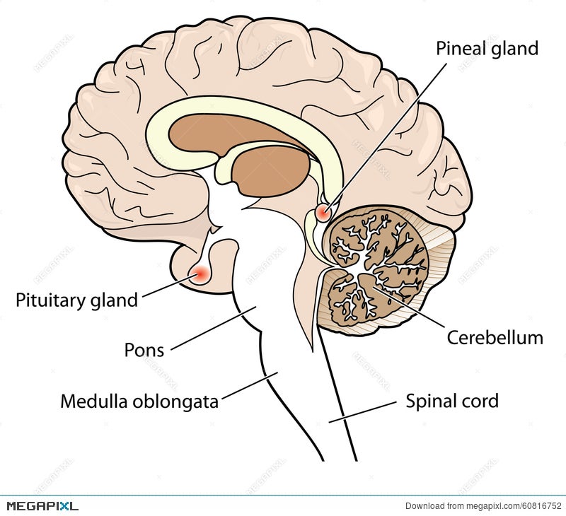
Pineal gland anatomical cross section vector illustration ...
human brain cross section diagram. 3d render, illustration ...
The brain - Macmillan Cancer Support

Lateral cross-section of the brain Diagram | Quizlet
Horizontal sections of the brain: Anatomy | Kenhub

About Brain Tumors - Dana-Farber Cancer Institute | Boston, MA

Human Brain Anatomy Diagram canvas print

The brain ion cross section showing the major structures and ...

interpreting a transverse section through brain

Cross Section Illustration Of Human Brain Showing Limbic ...

Brain Map 2

Human skull cross-section with brain, illustration - Stock ...
Brainstem Student notes
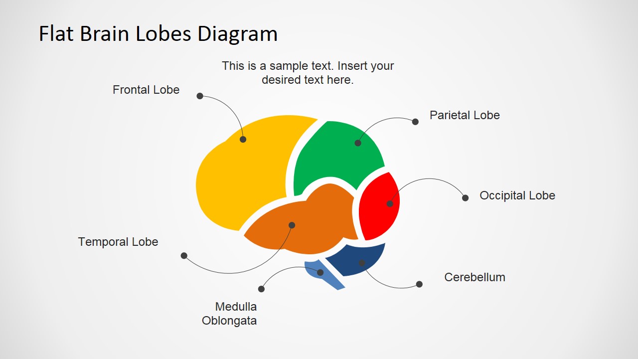
Flat Brain Lobes Cross Sectional Diagram for PowerPoint ...

AP Psych Test 3: Brain Cross-Section Diagram | Quizlet
Cross section brain Images, Stock Photos & Vectors | Shutterstock

human brain cross section model

Cross section of the human brain. | Download Scientific Diagram
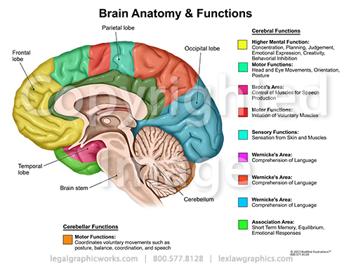
Brain Cross Sectional Anatomy & Functions - Legal Graphicworks
Brain Quiz | Project NEURON | University of Illinois

Brain Section Stock Illustrations – 1,730 Brain Section Stock ...
Cross Section of the Brain - Ocular Level

cross-section human brain 1 | Human brain anatomy, Human ...


Comments
Post a Comment