39 simple columnar epithelium labeled diagram
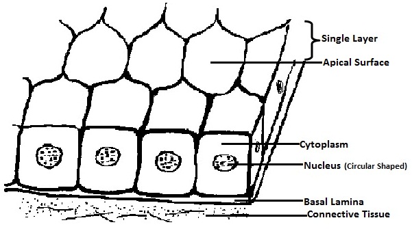
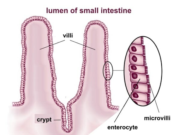
Ileum Histology Slide with Labeled Diagram and ... The simple columnar epithelium lines these villi of the ileum. ... I think the features mentioned earlier in the ileum histology slide labeled diagrams might help you a lot. Again, I will show you the ileum histology diagram where you might identify the following important features. ... Simple Columnar Epithelium |Introduction ,Types, & Functions The Simple Columnar Epithelium is tissues composed of a single layer of long epithelial cells which are usually seen in an area where absorption and secretion are important facts. The cells of these epithelial are arranged in a neat row within nuclei at the same level, near to th basal end.. In a cross-section of organs, these cells look like thin columns, which differentiate them from ...
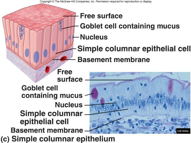
Simple Columnar Epithelium Labeled Diagram A simple columnar epithelium is a columnar epithelium that is uni-layered. In humans, a simple columnar epithelium lines most organs of the digestive tract including the stomach, . Simple Columnar Epithelium: A Labeled Diagram and Functions Epithelium is a tissue that lines the internal surface of the body, as well as the internal organs.

Simple columnar epithelium labeled diagram
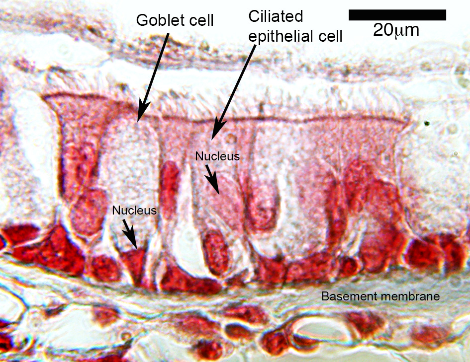
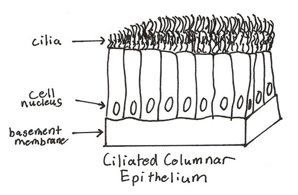
Simple Squamous Epithelium: Location and Diagram - Video ... The simple squamous epithelium is different from other types of epithelial tissue such as simple cuboidal, simple columnar, and stratified squamous epithelium in that it is only made of one layer ... PDF Marieb HA8 chapter 4 - Pearson Simple Cuboidal Epithelium (Figure 4.3b) Simple cuboidal epithelium consists of a single layer of cube-shaped cells. This epithelium forms the secretory cells of many glands, the walls of the smallest ducts of glands, and the walls of many tubules in the kidney. Its functions are the same as those of simple columnar epithelium. Simple columnar epithelium- structure, functions, examples 2. Ciliated columnar epithelium. The ciliated simple columnar epithelium is a single layer of ciliated column-like cells with oval nuclei near the base of cells, and the goblet cells are usually interspersed between the ciliated cells. The cilia are typically 5-10 μm long and 0.2 μm in diameter. Each cilium has a core structure consisting of nine peripheral microtubule doublets arrayed around by two central microtubules.
Simple columnar epithelium labeled diagram. Epithelium Diagram - Quizlet Form the Outer Covering of the skin and some internal organs. Form the Inner Lining of blood vessels, ducts and body cavities, and the interior of the respiratory, digestive, urinary and reproductive systems. Glandular epithelia. Constitute the secretory portion of glands. Simple squamous epithelium. Most delicate epithelium. 4.2 Epithelial Tissue - Anatomy & Physiology These epithelia are involved in the secretion and absorptions of molecules requiring active transport. Simple cuboidal epithelia are observed in the lining of the kidney tubules and in the ducts of glands. In simple columnar epithelium, the nucleus of the tall column-like cells tends to be elongated and located in the basal end of the cells. Like the cuboidal epithelia, this epithelium is active in the absorption and secretion of molecules using active transport. Epithelial Tissue - Anatomy and Physiology Like the cuboidal epithelia, this epithelium is active in the absorption and secretion of molecules. Simple columnar epithelium forms the lining of some sections of the digestive system and parts of the female reproductive tract. Ciliated columnar epithelium is composed of simple columnar epithelial cells with cilia on their apical surfaces. Simple epithelium: Location, function, structure | Kenhub Simple epithelium has only one cell layer where every cell is in direct contact with the underlying basement membrane. Generally, this type of epithelium is found inside the body probably due to the fragile nature and forms the lining of the body cavities, blood and lymph vessels, heart and respiratory system.. Being a thin layer has the physiological advantage of faster absorption and ...
Simple Columnar Epithelium - Kit Ng, Ph.D. Simple columnar epithelium contains cells that are rectangle in shape and have their nuclei arranged on the basal side. Their nuclei also tend to look oval shape. This type of epithelium can be found in the digestive tract, uterine tube and central canal of the spinal cord. In the digestive tract, structures called microvilli (or… PDF WEEK 1: COVERING AND LINING EPITHELIA - University of New ... These labelled diagrams should closely follow the current Science courses in histology, anatomy and embryology and complement the virtual microscopy used in the current Medical course. ... and simple columnar epithelium with basal striations FEATURE: lobe, lobules and ducts (TS) simple columnar epithelium | Histology slides, Tissue ... Columnar_Epithelial_Tissue :: A tissue that forms the linings of all internal and external body surfaces, such as the skin or the lining of the stomach, epithelial tissue is composed of layers of cells packed close together and tends to be thin.It has no blood vessels. Because of this, it depends on underlying tissues to provide it with the ... study.com › learn › lessonSimple Cuboidal Epithelium Function & Location | What Is ... Dec 13, 2021 · Simple Cuboidal Epithelium: Labeled Diagram Simple cuboidal epithelial cells are shaped like cubes, and the nucleus of each cell is large and located close to the center of the cell.
Structure-Function.org - Histology: Simple columnar ... Learn about the structures, locations, and functions of this epithelial tissue, and then test yourself with labeled images, hints, and answer keys that put you in control. Structure-Function.org structure-function About Contact Jejunum Histology Slide with Labeled Diagram and ... The lining epithelium of the tunica mucosa is a simple columnar epithelium with goblet cells. You may also find other different cells in the mucosa of the small intestine like penath cells, enteroendocrine cells, and others. The number of the plica circularis and villi mary varies in the different parts of an animal's small intestine. anatomylearner.com › epithelial-tissueEpithelial Tissue - Types, Location, Examples and Histology ... Apr 04, 2021 · Functions of simple columnar epithelium and their location. Absorption and secretion are the primary functions of simple columnar epithelium. You will find simple columnar epithelium in the following organs or structures of an animal’s body. #1. Simple ciliated columnar epithelium in the respiratory tract, uterine tube #2. Simple Columnar Epithelium - Definition & Function ... Simple columnar epithelia are tissues made of a single layer of long epithelial cells that are often seen in regions where absorption and secretion are important features. The cells of this epithelium are arranged in a neat row with the nuclei at the same level, near the basal end. In a cross-section of the organ, these cells appear like thin columns, differentiating them from flattened squamous cells and square-shaped cuboidal cells.
Simple Columnar Epithelium - an overview | ScienceDirect Topics
Answered: Under HPO, label the following from the… | bartleby Under HPO, label the following from the diagram of a sectioned colom showing the Simple columnar epithelium composed of secretory and columnar cells : goblet cells, columnar cells, cell membrane, cytoplasm, nucleus, lumen and basal lamina. fullscreen Expand.

3. Types of epithelial cells: simple squamous (top), simple ...
courses.lumenlearning.com › boundless-ap › chapterThe Male Reproductive System | Boundless Anatomy and Physiology The epithelium of the tubule consists of tall, columnar cells called Sertoli cells. Between the Sertoli cells are spermatogenic cells, which differentiate through meiosis to become sperm cells. There are two types of seminiferous tubules: convoluted, located toward the lateral side, and straight, as the tubule comes medially to form ducts that ...
Tissue Review Slides for Human Anatomy
Epithelium Lab - Yale University Simple columnar epithelium. Simple columnar epithelium consist of a single layer of cells that are taller than they are wide. This type of epithelia lines the small intestine where it absorbs nutrients from the lumen of the intestine. Simple columnar epithelia are also located in the stomach where it secretes acid, digestive enzymes and mucous.

simple columnar epithelium | Medical laboratory science ...
Ileum: Anatomy, histology, composition, functions | Kenhub The mucosa is lined by simple columnar epithelium (lamina epithelialis) comprising enterocytes and goblet cells. Underneath lies a connective tissue layer (lamina propria) and a muscle layer (lamina muscularis mucosae). Compared to the rest of the small intestine the circular folds are rather flat and the villi relatively short.
HLS [ Epithelial Tissue, Surface Specializations, and Glands ...
Simple Columnar Epithelium: A Labeled Diagram and ... Epithelium is a tissue that lines the internal surface of the body, as well as the internal organs. Simple epithelium is one of the types of epithelium that is divided into simple columnar epithelium, simple squamous epithelium, and simple cuboidal epithelium. Bodytomy provides a labeled diagram to help you understand the structure and function of simple columnar epithelium.
61 Simple Squamous Epithelium Illustrations & Clip Art - iStock
› 37006818 › Junqueiras_Basic(PDF) Junqueira's Basic Histology Text and Atlas, 14th ... Junqueira's Basic Histology Text and Atlas, 14th Edition

Describe Various Types of Epithelial Tissues with the Help of ...
Coursework Hero - We provide solutions to students We provide solutions to students. Please Use Our Service If You’re: Wishing for a unique insight into a subject matter for your subsequent individual research;
Epithelial Tissues Stock Illustrations – 30 Epithelial ...
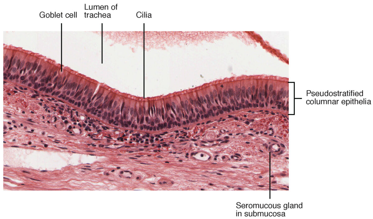
histology.medicine.umich.edu › resourcesConnective Tissue and Quiz 1 | histology Look at the areas outlined in the orientation diagram of the trachea and locate the loose, cellular connective tissue within the glands (the "glands" are coiled tubes of columnar epithelial cells; some the epithelial cells are tall and eosinophilic, whereas others are shorter and more basophilic).

draw well labelled diagrams of the following a columnar ...
Simple Columnar Epithelium Diagram - Quizlet Start studying Simple Columnar Epithelium. Learn vocabulary, terms, and more with flashcards, games, and other study tools.
Chapter 4 Tissues. Four Tissue Types Epithelia Connective ...
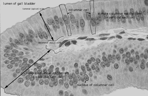
PDF Basic Histo diagrams labelled in colour - 2005 EPITHELIUM: simple squamous TISSUES / ORGANS: 1 endothelium lining blood vessel 2 mesothelium of serosa covering lung EPITHELIUM: simple columnar TISSUE / ORGAN: gall bladder EPITHELIUM: pseudostratified columnar (respiratory epithelium) MORE FULLY: pseudostratified ciliated columnar epithelium with goblet cells TISSUE / ORGAN: trachea EPITHELIUM: simple columnar or
Simple Cuboidal Epithelium
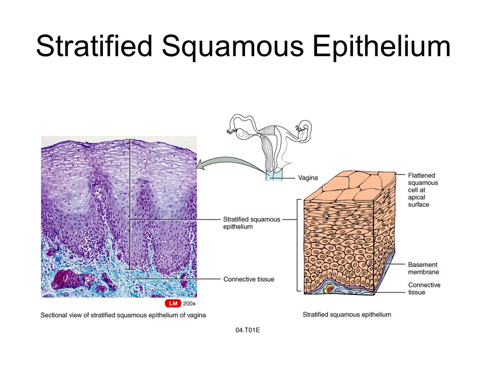
courses.lumenlearning.com › boundless-ap › chapterLayers of the Alimentary Canal | Boundless Anatomy and Physiology The most variation is seen in the epithelium tissue layer of the mucosa. In the esophagus, the epithelium is stratified, squamous, and non-keratinizing, for protective purposes. In the stomach. the epithelium is simple columnar, and is organized into gastric pits and glands to deal with secretion.
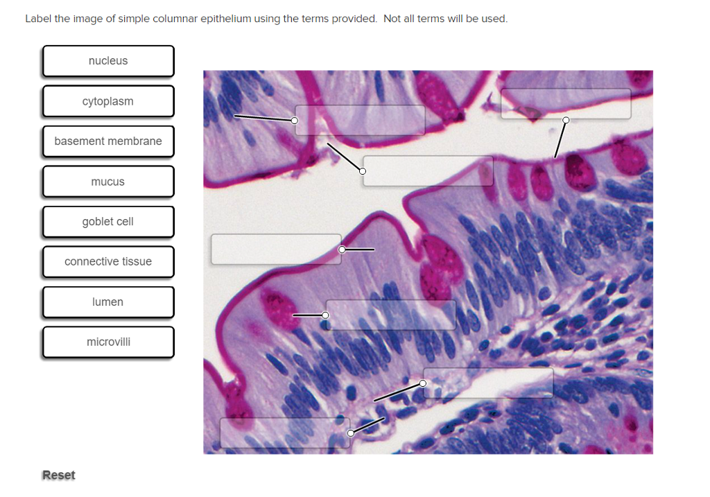
Solved Label the image of simple columnar epithelium using ...
› 44118315 › The_Biology_of_Cancer(PDF) The Biology of Cancer- R.Weinberg - Academia.edu Academia.edu is a platform for academics to share research papers.

PseudoStratified columnar epithelium | Psychology notes ...
Simple Columnar Epithelium Labeled Diagram Simple Columnar Epithelium: A Labeled Diagram and Functions Epithelium is a tissue that lines the internal surface of the body, as well as the internal organs. Simple epithelium is one of the types of epithelium that is divided into simple columnar epithelium, simple squamous epithelium, and simple cuboidal epithelium. Ciliated epithelium is a thin tissue that has hair-like structures on it. These hairs, called cilia, move back and forth to help move particles out of our body.

Epithelium Lab
BJC | The Beauty and Joy of Computing data:image/png;base64,iVBORw0KGgoAAAANSUhEUgAAAKAAAAB4CAYAAAB1ovlvAAAAAXNSR0IArs4c6QAAArNJREFUeF7t1zFqKlEAhtEbTe8CXJO1YBFtXEd2lE24G+1FBZmH6VIkxSv8QM5UFgM ...
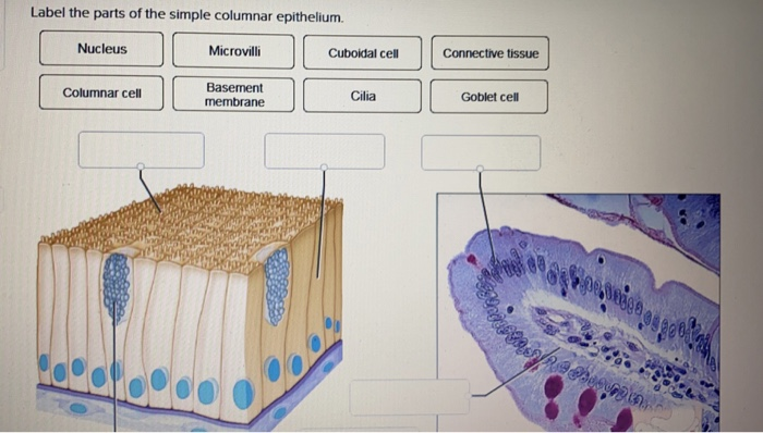
Solved Label the parts of the simple columnar epithelium ...
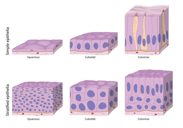
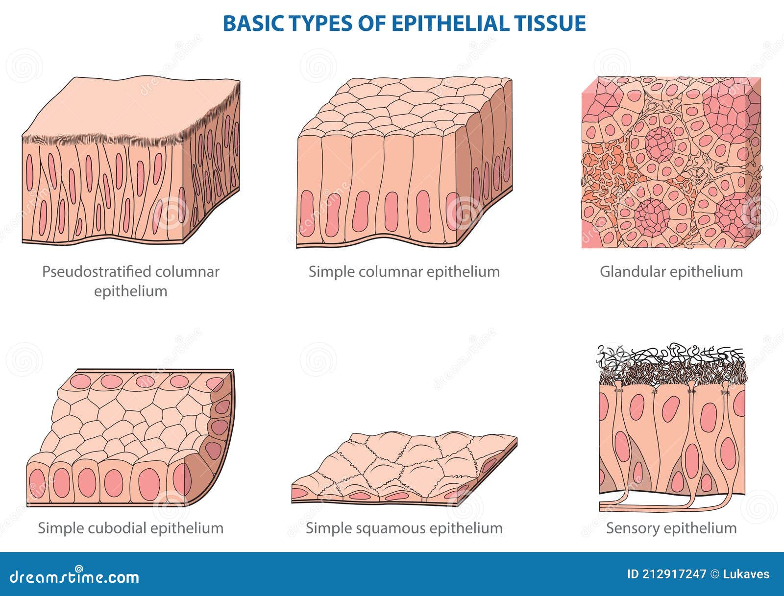
Epithelial Tissue: Structure with Diagram, Function, Types ... Simple Epithelium- it is composed of one layer of a cell and mostly has a secretory or an absorptive function. Compound (Stratified) Epithelium- it is made up of two or more than two layers of cells and mostly has a protective function. The glandular epithelium is made up of cuboidal or columnar cells. They are specialised for secretion.

Epithelial Tissues Lab – David Fankhauser
Simple columnar epithelium - Eugraph These absorptive cells are a single layer of columnar cells. (a simple columnar epithelium). Note an oval nucleus in the lower part of each columnar cell. Arrows indicate the base of this simple columnar epithelium sce. The lumen is indicated by lu. The surface area for absorption is increased by projections of the intestinal wall called villi.

4.2 Epithelial Tissue – Anatomy & Physiology
Simple columnar epithelium- structure, functions, examples 2. Ciliated columnar epithelium. The ciliated simple columnar epithelium is a single layer of ciliated column-like cells with oval nuclei near the base of cells, and the goblet cells are usually interspersed between the ciliated cells. The cilia are typically 5-10 μm long and 0.2 μm in diameter. Each cilium has a core structure consisting of nine peripheral microtubule doublets arrayed around by two central microtubules.

How to draw stratified columnar epithelium || easy way
PDF Marieb HA8 chapter 4 - Pearson Simple Cuboidal Epithelium (Figure 4.3b) Simple cuboidal epithelium consists of a single layer of cube-shaped cells. This epithelium forms the secretory cells of many glands, the walls of the smallest ducts of glands, and the walls of many tubules in the kidney. Its functions are the same as those of simple columnar epithelium.

Stratified Cuboidal Epithelium Diagram | Quizlet
Simple Squamous Epithelium: Location and Diagram - Video ... The simple squamous epithelium is different from other types of epithelial tissue such as simple cuboidal, simple columnar, and stratified squamous epithelium in that it is only made of one layer ...
Epithelia: The Histology Guide

Gallbladder Simple Columnar Epithelium Stock Photo - Download ...

Study Notes
Epithelium
Surface Epithelium: Histology | Concise Medical Knowledge

Simple Squamous Epithelium 40X - Annotated | Histology
What is the epithelial tissue? - Quora
Simple Columnar Epithelium - Definition & Function | Biology ...

Simple Squamous Epithelium Diagram | Quizlet
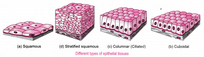
Describe the structure and function of different types of ...
Ciliated Columnar Epithelium

Simple Squamous Epithelium
ANIMAL TISSUE — Biology Notes

Pseudostratified Columnar Epithelium Function & Location ...

Study Notes
Epithelia: The Histology Guide

Simple Columnar Epithelium Diagram | Quizlet

Simple Columnar Epithelium, Stomach Pyloric, 400x | Histology
Simple epithelium: Location, function, structure | Kenhub
Ch. 4


Comments
Post a Comment