42 nerves of the lower limb diagram
Cutaneous nerves of lower limb Diagram | Quizlet Start studying Cutaneous nerves of lower limb. Learn vocabulary, terms, and more with flashcards, games, and other study tools. Anatomy Tables - Muscles of the Lower Limb posterior division of the obturator nerve; tibial nerve (ischiocondylar part) obturator a., deep femoral a., medial femoral circumflex a. the ischiocondylar part of adductor magnus is a hamstring muscle by embryonic origin and action, so it is innervated by the tibial nerve: adductor minimus: lower portion of the inferior pubic ramus
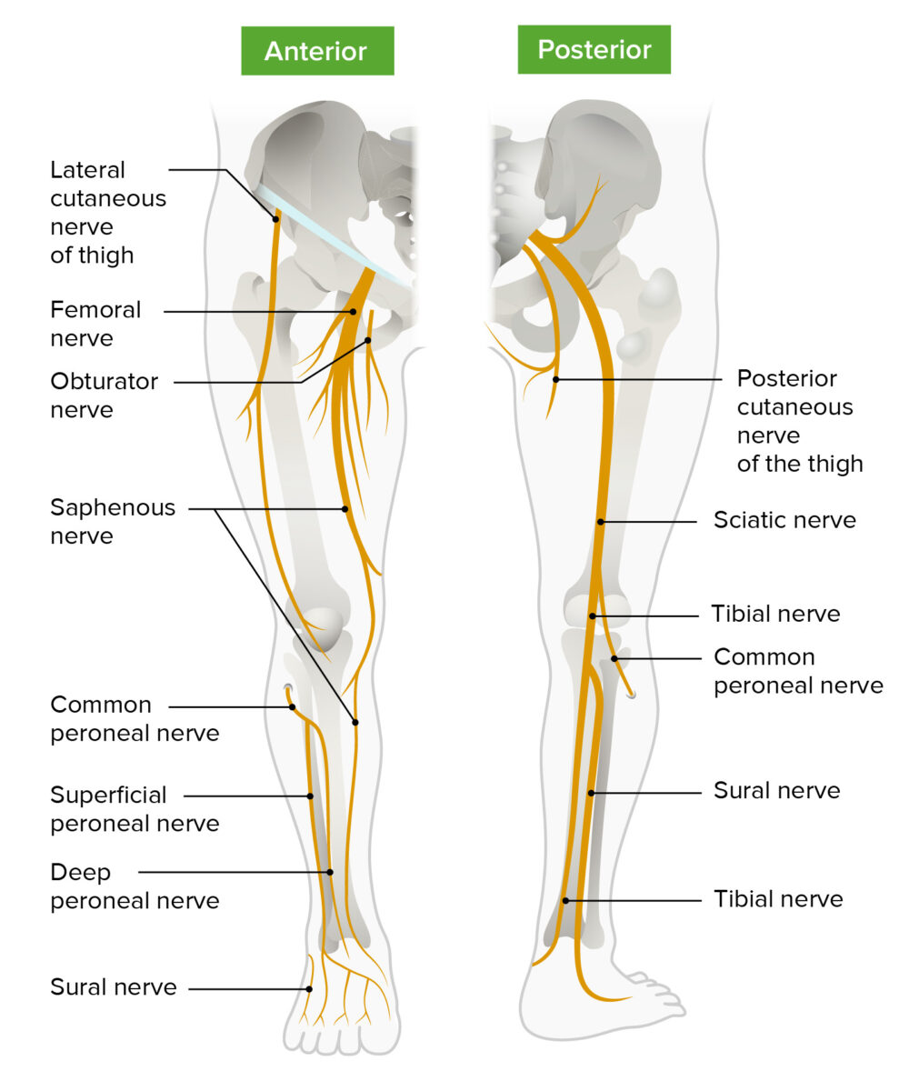
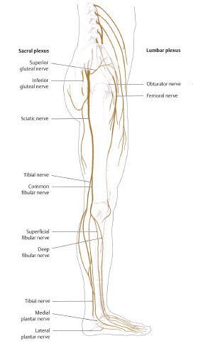
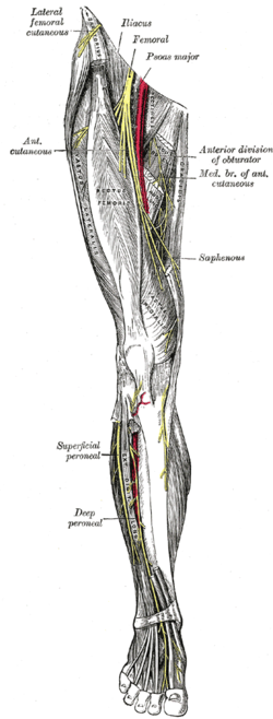
Lower limb arteries and nerves: Anatomy, branches | Kenhub It originates from the sacral plexus (L4-S3) and travels all the way down the posterior aspect of the lower limb., The sciatic nerve innervates the entire skin of the leg, the posterior thigh muscles, and the muscles of the leg and foot. The obturator nerve innervates the adductor muscles as well as the skin on the medial aspect of the thigh.
Nerves of the lower limb diagram
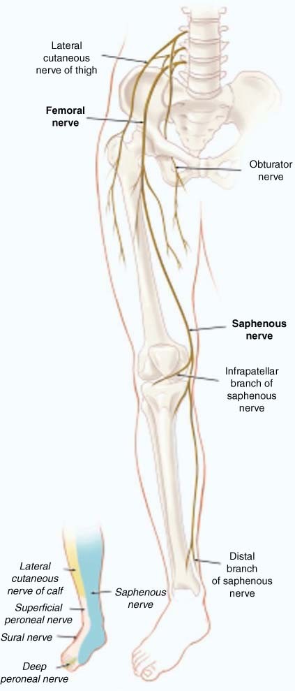
nerves of the leg diagram - ModernHeal.com This image is titled nerves of the leg diagram and is attached to our article about Leg Nerves and Reflex Motion in Feet.. Be sure to visit the guide for more context and information about nerves of the leg diagram, or read some of our other Health & Anatomy posts! Nerves of the Leg and Foot | Interactive Anatomy Guide The lumbar plexus forms in the lower back from the merger of spinal nerves L1 through L4 while the sacral plexus forms in the pelvic region from spinal nerves L4, L5, and S1 through S4. The femoral, saphenous, obturator, and lateral femoral cutaneous nerves all extend from the lumbar plexus into the muscles and skin of the thigh and leg. Cranial Nerves Summary | Anatomy | Geeky Medics Oct 22, 2021 · Oculomotor nerve (CNIII) CN III is the oculomotor nerve.It provides general somatic efferent and general visceral efferent fibres to the extraocular muscles and pupillary constrictor muscles respectively. It is the efferent limb for the pupillary light reflex. The muscles are the levator palpebrae superioris, inferior oblique, and superior, medial and inferior recti.
Nerves of the lower limb diagram. Peripheral Nerves and Arteries of the Lower Extremity ... Peripheral Nerves and Arteries of the Lower Extremity. Neurological Assessment of the Distal Lower Extremity. Sensory - as per cutaneous innervation in side diagrams. Superficial Peroneal Nerve: ask the patient to evert the foot. Deep Peroneal Nerve: ask the patient to dorsi flex the foot (L4) and extend the toes (L5) Tibial Nerve: ask the ... Spinal Nerve Chart - Miller Chiropractic Clinic L1-L5 is the LOWER BACK. Simply line up the "Vertebral Level" with the "Possible Symptoms" and you will see some surprising connections of symptoms that relate to your spine. Chiropractic is great for back, neck and extremity pain. It is also great for restoring proper nerve function to the rest of your body that you don't think about. Nerve Injuries of the Lower Extremity | Clinical Gate In a recent review of lower extremity nerve injuries in Wroclaw, Poland, 1 the incidence of lower extremity nerve injuries was 20% of upper extremity nerve injuries. Irrespective of series, the peroneal nerve was the single most common lower extremity nerve to be injured. All the mechanisms of nerve damage, stretch, contusion, laceration, and ... Solved Part A Drag the labels onto the diagram to identify ... Anatomy and Physiology questions and answers. Part A Drag the labels onto the diagram to identify the nerves of the lower trunk and lower limb. Reset Help Intonoglutoal otvo Femoral nerve Letcalfemoral cutaneous nerve HINDI Scalerave Pucend nerve llicingumal nerve Sapronoue verve Submit Request Answer. Question: Part A Drag the labels onto the ...
The Brachial Plexus - Sections - Branches - TeachMeAnatomy The brachial plexus is a network of nerve fibres that supplies the skin and musculature of the upper limb. It begins in the root of the neck, passes through the axilla, and runs through the entire upper extremity. The plexus is formed by the anterior rami (divisions) of cervical spinal nerves C5, C6, C7 and C8, and the first thoracic spinal nerve, T1. Intercostal nerves - Wikipedia The first two nerves supply fibers to the upper limb and thorax; the next four distribute to the walls of the thorax; the lower five supply the walls of the thorax and abdomen. The 7th intercostal nerve end at the xyphoid process of the sternum. The 10th intercostal nerve terminates at … Lower subscapular nerve - Wikipedia The lower subscapular nerve contains axons from the ventral rami of the C5 and C6 cervical spinal nerves. It is the third branch of the posterior cord of the brachial plexus. It gives branches to 2 muscles: subscapularis muscle. It usually gives 4 branches to innervate the subscapularis, and can give up to 8 branches. teres major muscle. Dermatomes of the lower limb. | Download Scientific Diagram Download scientific diagram | Dermatomes of the lower limb. from publication: Physiotherapy management of sciatica | Sciatica, Radiculopathy and Low Back Pain | ResearchGate, the professional ...
Nerves of Lower Limb - Color Diagram Nerves of Lower Limb - Color Diagram. High Resolution Poster. Click Here. Citing Research References. When you research information you must cite the reference. Citing for websites is different from citing from books, magazines and periodicals. The style of citing shown here is from the MLA Style Citations (Modern Language Association). Anatomy of lower extremity - eAnatomy - IMAIOS A diagram shows the various inguinal lymph nodes (lymphatic ganglia). The chapter on the innervation of the lower limb presents diagrams of the lumbosacral plexus and its main nerve branches for the lower limb (lateral cutaneous nerve of the thigh, femoral nerve, sciatic nerve and posterior cutaneous nerve of the thigh and obturator nerve). Lower Back Skeleton Photos and Premium High ... - Getty Images Illustration of lower limb, skeletal muscles, front and back views. the bones of the lower back - lower back skeleton stock illustrations. ... the nerves of the lower back - lower back skeleton stock illustrations. the nerves of the lower back - lower back skeleton stock illustrations. lecture 26 Nerves of Lower limb Diagram | Quizlet Start studying lecture 26 Nerves of Lower limb. Learn vocabulary, terms, and more with flashcards, games, and other study tools.
Lumbosacral plexus and innervation of lower limb – Human ...
Nerves Of The Lower Leg 3D - Everything You Need To Know ... Dr. Ebraheim's educational animated video showing 3D animation of the nerves of the lower leg.The tibial nerve is a branch of the sciatic nerve. The tibial n...

Lower Limb Nerves 1 Quiz
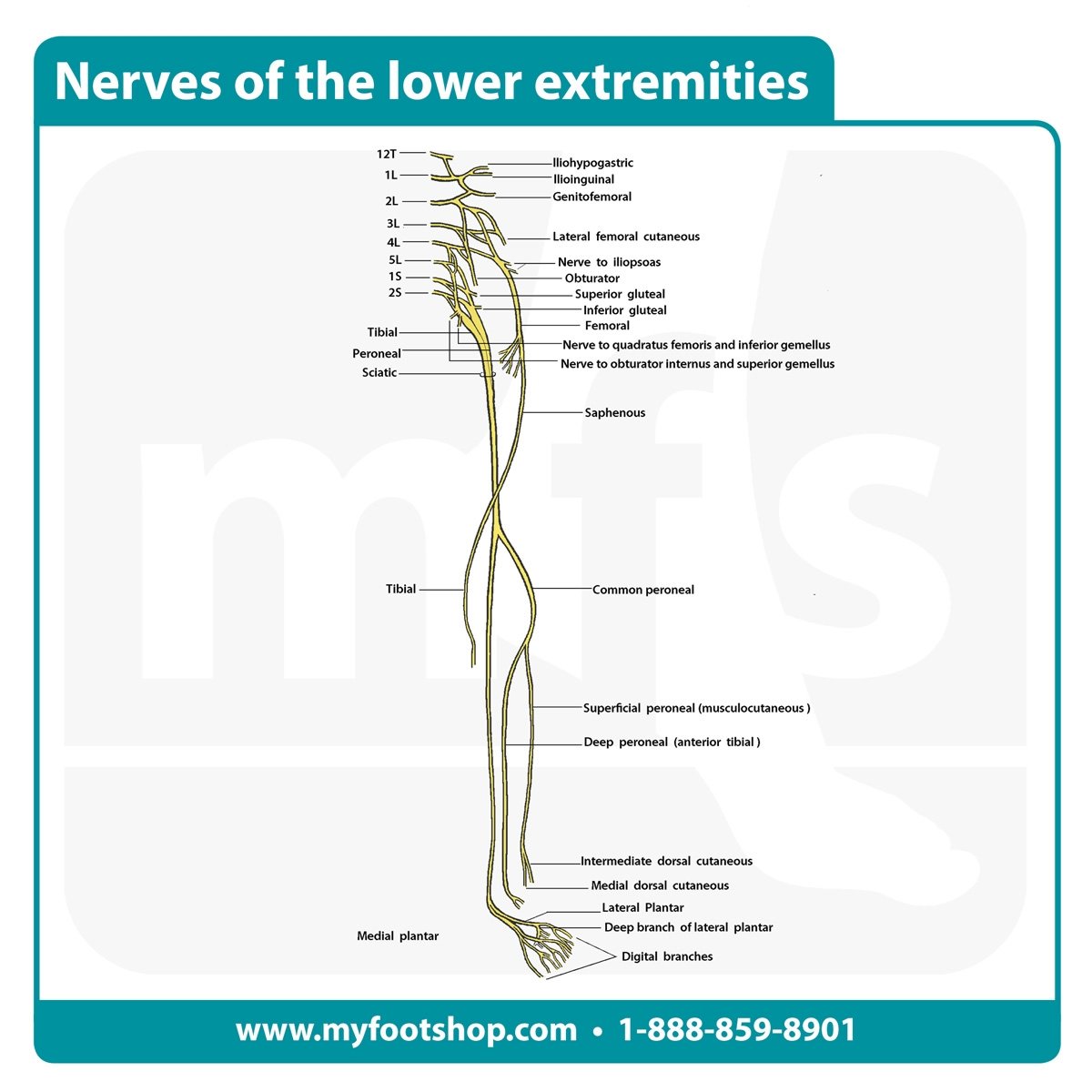
Nerve supply of the lower limb - Musculoskeletal Key The first lumbar nerve divides into two, the iliohypogastric and the ilioinguinal nerves, which supply the skin of the buttock and groin, respectively. A third cutaneous nerve, the genitofemoral, is formed from L1 and L2, and supplies a small area of skin on the upper front part of the thigh. Sacral plexus: position and formation

Sacral plexus (diagram) | Image | Radiopaedia.org
Nerves of the Abdomen, Lower Back and Pelvis - Innerbody The nervous system of the abdomen, lower back, and pelvis contains many important nerve conduits that service this region of the body as well as the lower limbs. This section of the nervous system features the most inferior portion of the spinal cord along with many major nerves, plexuses, and ganglia that serve the vital organs of the ...
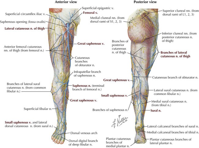
Cutaneous Innervation of the Lower Limb
Anatomy, Appendicular Skeleton - StatPearls - NCBI Bookshelf Jul 31, 2021 · The lymphatics of the upper and lower limb primarily follow the major blood vessels. Nerves. The upper extremity nerves originate from the brachial plexus. The brachial plexus is composed of roots, trunks, divisions, cords, and ultimately the five named branches. Spinal nerve roots C5 to T1 contribute to the brachial plexus.

Anatomical review of the lumbosacral plexus and nerves of the ...
Nerves of the Upper Limb - TeachMeAnatomy There are 6 topics covered in the nerves of the upper limb, an overview of the brachial plexus and a more in-depth look into it's 5 main branches: axillary, musculocutaneous, median, radial, and ulnar nerves.. The brachial plexus is a collection of nerve fibres that supply motor and sensory innervation to the upper limb. It originates from nerve roots C5 to T1 and, as it passes through the ...
.png)
Cambridge Questions
Lower Limb - 3D Interactive Anatomy Tutorials 3D interactive models and tutorials on the anatomy of the lower limb, including the muscular compartments, osseus structures, blood supply and innervation.

Robotic Leg Control with EMG Decoding in an Amputee with ...
Leg: Anatomy and Function of Bones and Muscles, Plus Diagram Jun 18, 2018 · The lower leg extends from the knee to the ankle. This area is commonly referred to as the calf. Lower leg bones. Tibia. Also called the shin bone, the tibia is the longer of the two bones in the ...
Lower limb arteries and nerves: Anatomy, branches | Kenhub
Sensory cranial nerves: Anatomy, functions and diagram ... Feb 17, 2022 · The optic nerves are additionally covered by extensions of the meninges which cover the brain. The optic nerves enter the cranial cavity via the optic canals, and enter the brain at the pre-optic region of the diencephalon. Upon entering the brain, the optic nerves join to form an “x”-shaped structure called the optic chiasm. It is here ...
![Figure, Back of left lower extremity,...] - StatPearls - NCBI ...](https://www.ncbi.nlm.nih.gov/books/NBK546623/bin/Gray1247.jpg)
Figure, Back of left lower extremity,...] - StatPearls - NCBI ...
Lumbar Spinal Nerves - Spine-health L2, L3, and L4 spinal nerves provide sensation to the front part of the thigh and inner side of the lower leg. These nerves also control movements of the hip and knee muscles. L5 spinal nerve provides sensation to the outer side of the lower leg, the upper part of the foot, and the web-space between the first and second toe.
Lower Limb – Earth's Lab
Inferior cluneal nerves | Psychology Wiki | Fandom Inferior cluneal nerves. Diagram of the segmental distribution of the cutaneous nerves of the right lower extremity. Posterior view. The inferior clunial nerves innervate the skin of the lower part of the buttocks. They arise as branches of the posterior cutaneous nerve of the thigh .

File:2136ab Lower Limb Veins Anterior Posterior.jpg - Wikipedia
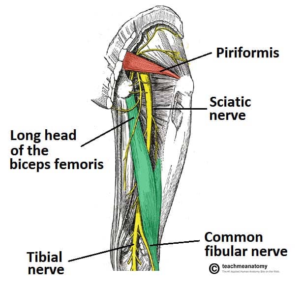
Nerves of the Lower Limb - TeachMeAnatomy Nerves of the Lower Limb; The Lumbar Plexus. View Article. The Sacral Plexus. View Article. The Femoral Nerve. View Article. The Obturator Nerve. View Article. The Sciatic Nerve. View Article. Tibial Nerve. View Article. The Common Fibular Nerve. View Article. The Superficial Fibular Nerve. View Article.
Nerve Entrapments of the Lower Leg, Ankle and Foot in Sport ...
Lower Leg Anatomy, Diagram & Pictures | Body Maps The main muscle in this area of the leg is the gastrocnemius, which gives the calf a bulging muscular appearance. Some nerves of the sacral plexus innervate this area, namely the superficial...

The nerve supply of the lower limb, Stock Photo, Picture And ...
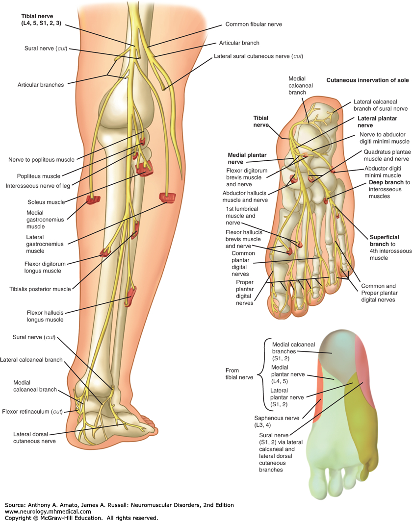
The Tibial Nerve - Course - Motor - Sensory - TeachMeAnatomy Jan 19, 2021 · The tibial nerve is a major peripheral nerve of the lower limb. It has several cutaneous and motor functions in the leg and foot. In this article, we shall look at the anatomy of the tibial nerve – its anatomical course, functions and clinical correlations.

8 Lower limb nerve ideas | lower limb, limb, nerve
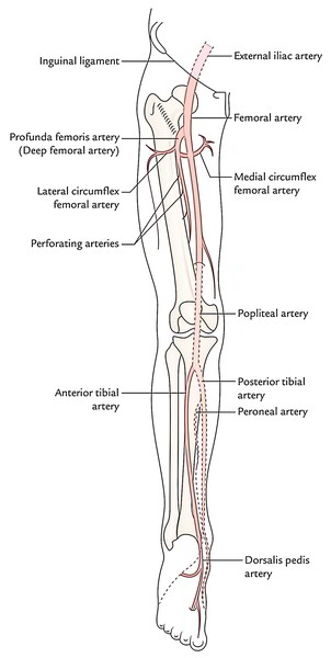
Major arteries, veins and nerves of the body: Anatomy | Kenhub Feb 14, 2022 · Arteries, veins and nerves of the lower limb (diagram) The main artery of the lower limb is the femoral artery and its continuation–the popliteal artery. The femoral artery supplies the gluteal region and the thigh before it continues as the popliteal artery in the posterior knee.The popliteal artery then supplies the knee region, before splitting into two branches which supply the leg ...
Leg | Concise Medical Knowledge
PDF Dermatomes Anatomy Overview between the peripheral nerves of the limbs (upper and lower extremities) is far less extensive (see the following image). Thus, in the limbs, complete interruption of a single peripheral nerve typically produces changes in sensation that are, indeed, appreciated by a patient. Dermatomes of the extremities.
Lower Limb | Basicmedical Key
PDF Lower Limb - Lippincott Williams & Wilkins The lower limb is designed for weight-bearing, balance, and mobility. The bones and muscles of the lower limb are larger and stronger than those of the upper limb, which is necessary for the functions of weight-bearing and balance. Our lower limbs carry us, allow us to push forward, and also keep us standing still.

Nerves of the lower extremity - UpToDate
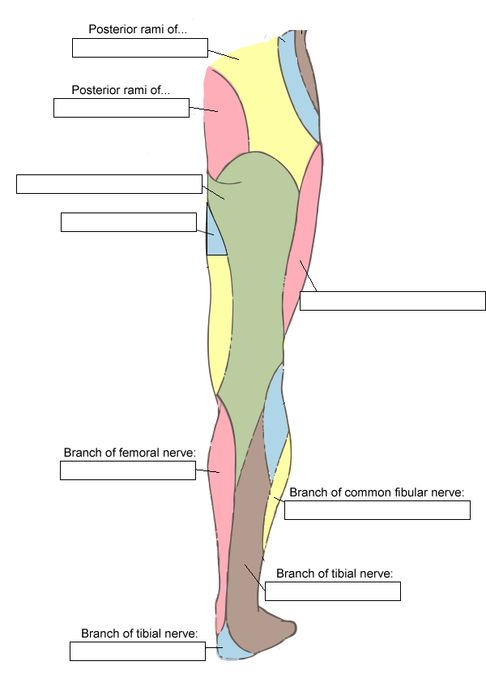
Cutaneous innervation in the lower limb. | Download ... The main nerves in the knee region are the tibial nerve, the common peroneal nerve, and the saphenous nerve. These three nerves innervate the lower leg and foot, providing sensory and motor...

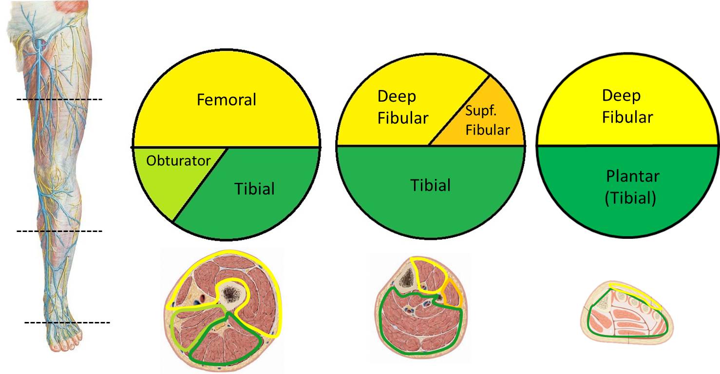
Anatomical landmarks used for obtaining CSA measurements for ...
PDF Summrry for Main Nerves of The Upper Limb NERVES OF THE UPPER LIMB : nerve Course branches Injuries The Axillary Nerve * arises from the posterior cord of the brachial plexus (C5 and 6), * This nerve passes to the posterior aspect of the arm through the quadrangular space in the company of the posterior circumflex vessels. * On emerging from the quadrangular space, the
Nerves of the Lower Limb - Labeling Diagram
Solved Drag the labels onto the diagram to identify the ... Transcribed image text: Drag the labels onto the diagram to identify the anatomical landmarks of the spinal cord. Reset Help Dura mater Posterior median BU Control canal Anterior view of spinal cord Voltool Anterior y hom II III La gray hom Posterior any hom Anboto median Donin rool ganglion Submit Request Answer Label the nerves of the lower trunk and lower limb.
Nerves of the Lower Limb - TeachMeAnatomy
Nerve supply of the human leg - Wikipedia Nerve supply of the human leg. Lower limb. Foot. Cutaneous innervation refers to the area of the skin which is supplied by a specific nerve . Modern texts are in agreement about which areas of the skin are served by which nerves, but there are minor variations in some of the details. The borders designated by the diagrams in the 1918 edition of ...
Public Home - AENS
Lower limb anatomy: Bones, muscles, nerves, vessels | Kenhub In terms of innervation, the leg receives it via the common fibular/peroneal, tibial, and saphenous nerves. The first two are branches of the sciatic nerve while the latter stems from the femoral nerve. These three nerves divide further to supply the various structures of the leg. Neurovasculature of the leg and knee Explore study unit
Lumbosacral plexus and innervation of lower limb – Human ...
Larry M - Yavapai College Diagram and follow the path of spinal nerves through the lumbral/sacral plexus out to the muscles of the upper limb. Diagram the path of sensory innervation from regions of the upper limb through the major nerves to the spinal cord; Predict the type of condition/impairment caused by nerve damage to major nerves of the lower limb;

Medicine as a profession - Nerves Anatomy Of The Lower Limb ...
Cranial Nerves Summary | Anatomy | Geeky Medics Oct 22, 2021 · Oculomotor nerve (CNIII) CN III is the oculomotor nerve.It provides general somatic efferent and general visceral efferent fibres to the extraocular muscles and pupillary constrictor muscles respectively. It is the efferent limb for the pupillary light reflex. The muscles are the levator palpebrae superioris, inferior oblique, and superior, medial and inferior recti.

Instant Anatomy - Lower Limb - Vessels - Arteries - General ...
Nerves of the Leg and Foot | Interactive Anatomy Guide The lumbar plexus forms in the lower back from the merger of spinal nerves L1 through L4 while the sacral plexus forms in the pelvic region from spinal nerves L4, L5, and S1 through S4. The femoral, saphenous, obturator, and lateral femoral cutaneous nerves all extend from the lumbar plexus into the muscles and skin of the thigh and leg.

Nerve Innervation of Upper and Lower Extremities
nerves of the leg diagram - ModernHeal.com This image is titled nerves of the leg diagram and is attached to our article about Leg Nerves and Reflex Motion in Feet.. Be sure to visit the guide for more context and information about nerves of the leg diagram, or read some of our other Health & Anatomy posts!
Nerves of the lower extremity | Lower extremity anatomy ...

Nerve Supply of the Lower Limb - ppt video online download

Prac 6, Nerves of Lower Limb Diagram | Quizlet
Saphenous nerve - Wikipedia

Nerves to posterior lower limb

lecture 26 Nerves of Lower limb Diagram | Quizlet
Cutaneous nerves of the lower limb (posterior) - KwizMi Medical

8 Lower limb nerve ideas | lower limb, limb, nerve
1 1 Anatomy – Lower limb – Nerves, Vessels, Lymphatics

Clinical Anatomy of Lower Extremity | Basicmedical Key

Nerves of the lower limb – Mednotes.

Osseous tissue and dermatome innervation of the lower ...

Vessels & nerves of the lower limb Diagram | Quizlet
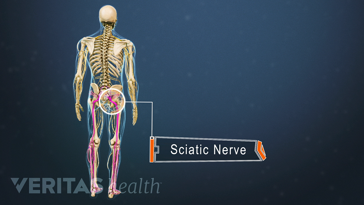
Sciatic Nerve Anatomy

Amino Neuro Frequency Therapy - ANF Therapy ...
![PDF] Lower Extremity Nerve Entrapments in Athletes | Semantic ...](https://d3i71xaburhd42.cloudfront.net/bc50b88bed00e16bdb31594ba1c34fd572a86918/2-Figure1-1.png)
PDF] Lower Extremity Nerve Entrapments in Athletes | Semantic ...

M6040 - LOWER LIMB NERVES ANTERIOR


Comments
Post a Comment