40 leg veins diagram
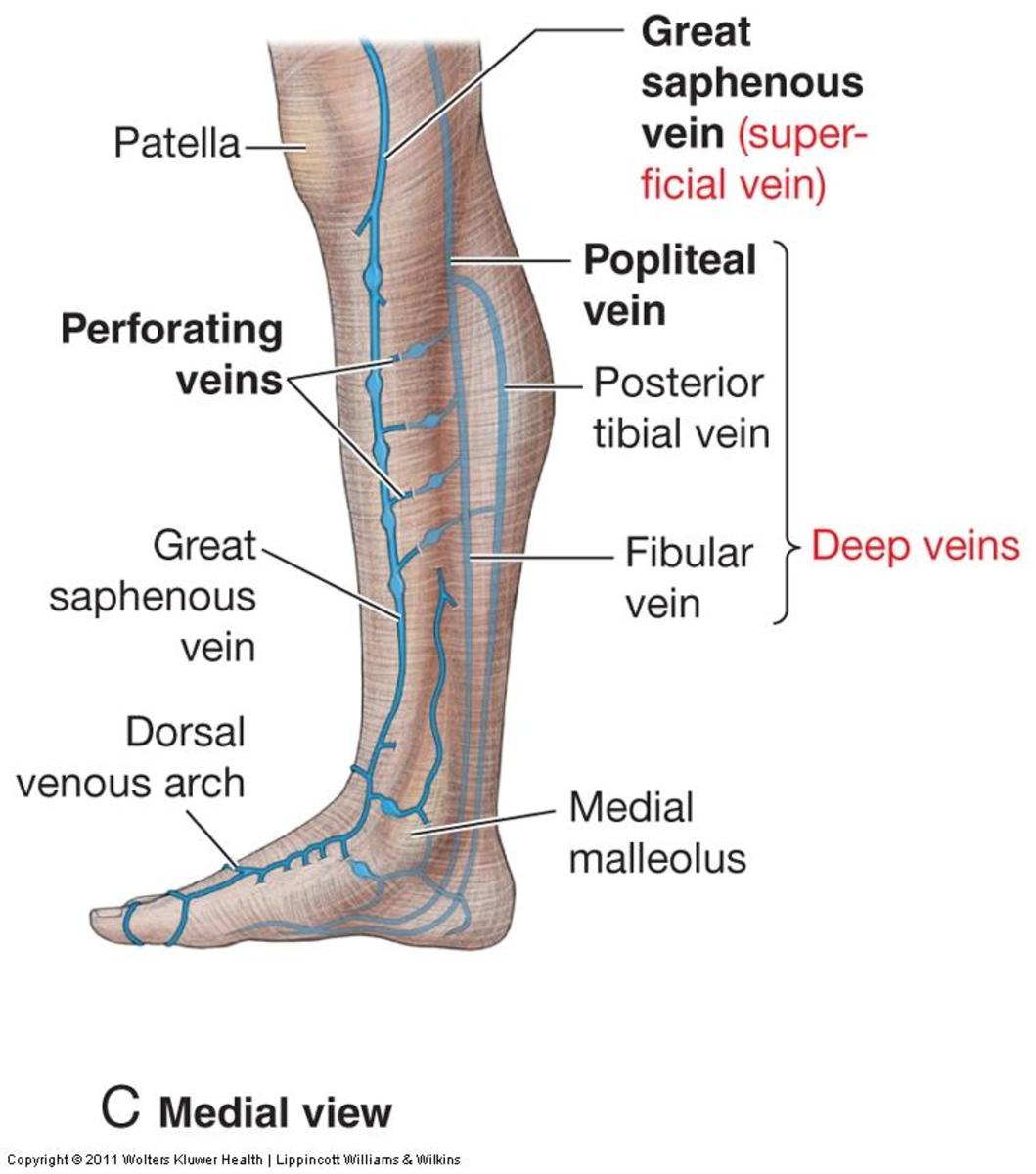
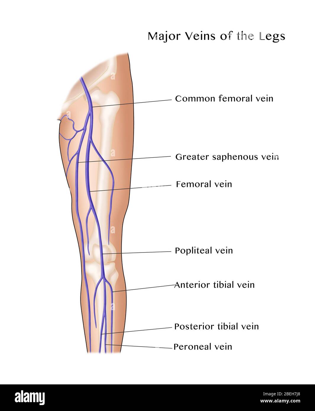
Veins (in blue) are the blood vessels that return blood to the heart. Deep veins, located in the center of the leg near the leg bones, are enclosed by muscle. The iliac, femoral, popliteal and tibial (calf) veins are the deep veins in the legs. Superficial veins are located near the surface of the skin and have very little muscle support. Bird Leg Anatomy - Bones, Muscles, Sciatic Nerve, and Shank Vein with Labeled Diagram 14/12/2021 16/10/2021 by anatomylearner The bird leg anatomy exhibits several specialized features compared to that of mammals. They are using for weight-bearing, scratching, climbing, grasping, and swimming.
2,217 leg veins anatomy stock photos, vectors, and illustrations are available royalty-free. See leg veins anatomy stock video clips. of 23. leg veins leg vein lower limb anatomy medical anatomy veins legs arterial leg veins anatomy leg ulceration veins legs saphenous vein superficial veins of the leg. Try these curated collections.

Leg veins diagram
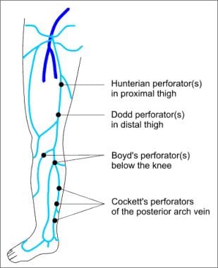
These veins range in number from 7 to 22, averaging ∼14 per leg, with 52% being direct perforators. 11 The lower perforators tend to be posterior tibial perforators, and the more superior perforators are paratibial. 11 Inferior perforators tend to be short, often only 1 cm in length, whereas toward the middle of the leg they may be 3 to 4 cm ... Arteries and Veins of the Leg. Create healthcare diagrams like this example called Arteries and Veins of the Leg in minutes with SmartDraw. SmartDraw includes 1000s of professional healthcare and anatomy chart templates that you can modify and make your own. 14/71 EXAMPLES. EDIT THIS EXAMPLE. CLICK TO EDIT THIS EXAMPLE. the vein along the posteromedial aspect of the fibula is the peroneal vein. In a US approach, the cortical shadow of the tibia and fibula can be used as a bony landmark. The paired veins are present on both sides of the artery (Fig. 4F). After stretching the patient's leg, the anterior tibial vein can be visualized from an anterolateral
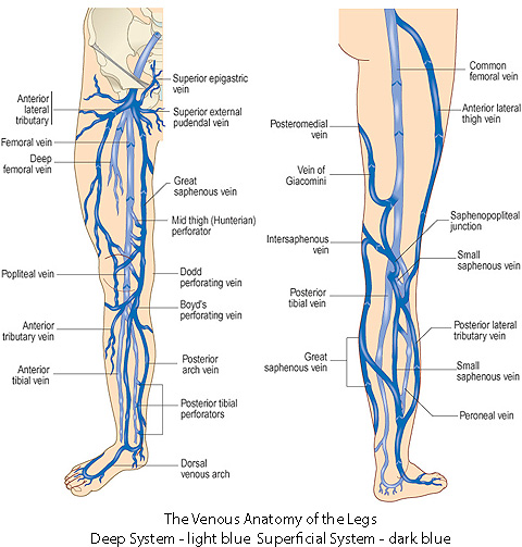
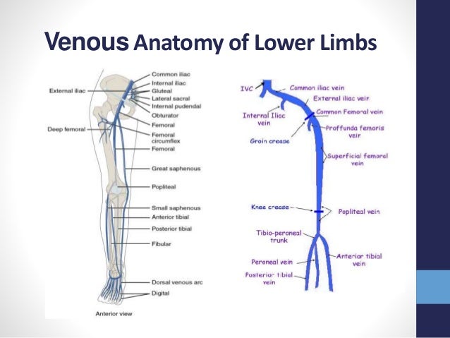
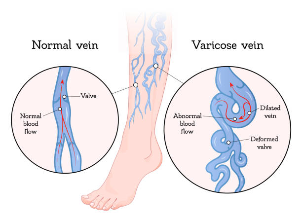
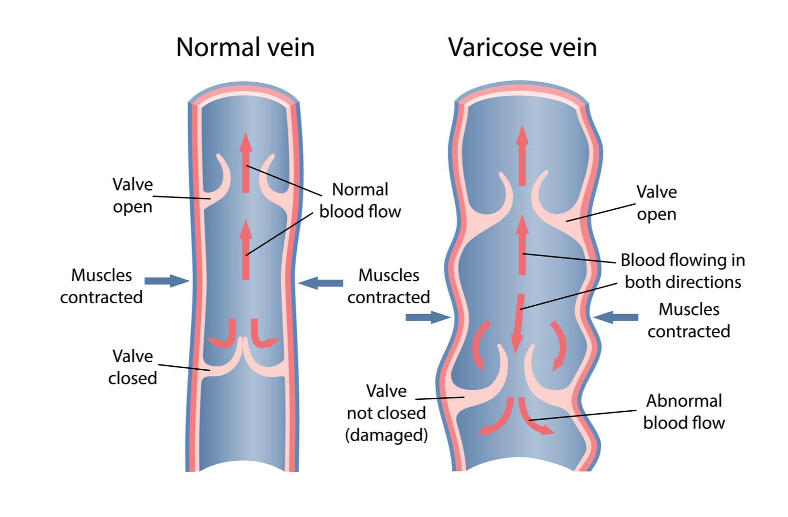
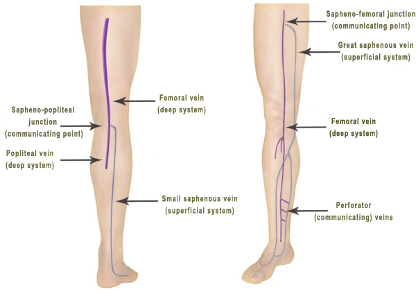
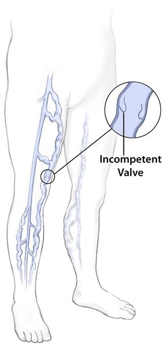
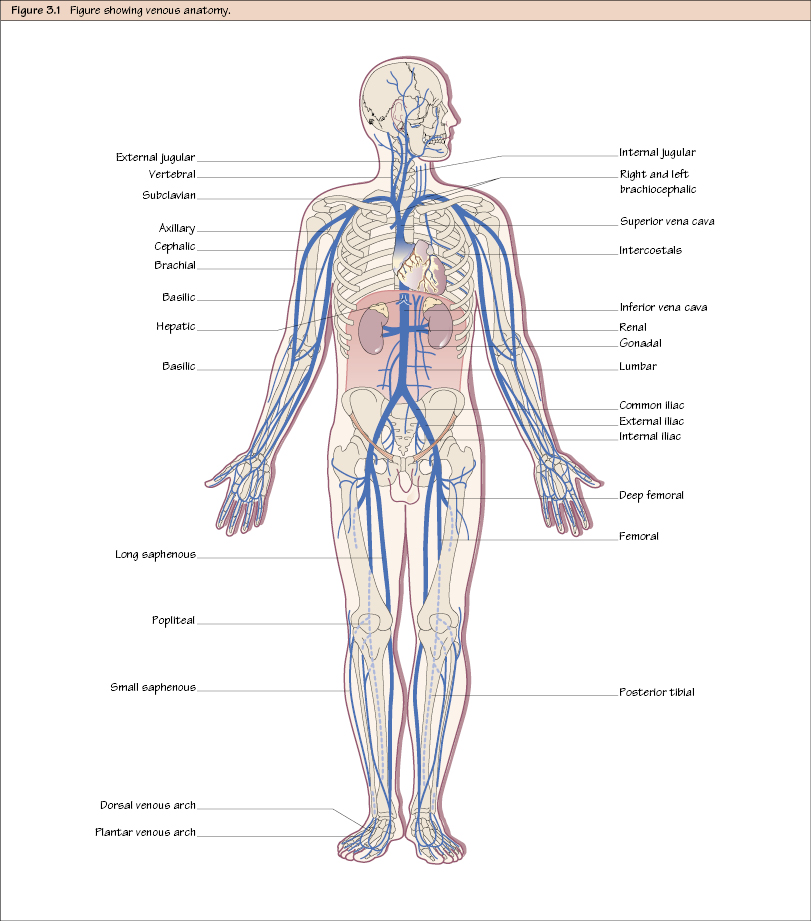
Leg veins diagram. Basilic vein . Brachial vein . Median cubital vein . Dorsal venous arch . Small saphenous vein . Digital veins . Great saphenous vein . Ulnar vein . Femoral vein . External iliac vein . Popliteal vein . Anterior tibial vein . Posterior tibial vein . Subclavian vein . Cephalic vein . Dorsal metatarsal veins The long saphenous vein has many connections with the short saphenous vein and the deep veins of the lower limb via perforating veins.Just distal to the knee, the long saphenous vein communicates and receives blood from the small saphenous vein, anterior and posterior tibial veins.The main tributaries of the long saphenous vein join it in the thigh, near its junction with the femoral vein. Start studying leg veins. Learn vocabulary, terms, and more with flashcards, games, and other study tools. Leg Vein Problems. Veins carry deoxygenated (oxygen deficient) blood and waste products back to the liver, heart and lungs. If the blood flow to the trunk is impeded then the circulation in the legs becomes sluggish (refer to the diagram below). This may occur due to : Valve incompetence (varicose veins) which affects the superficial leg veins.
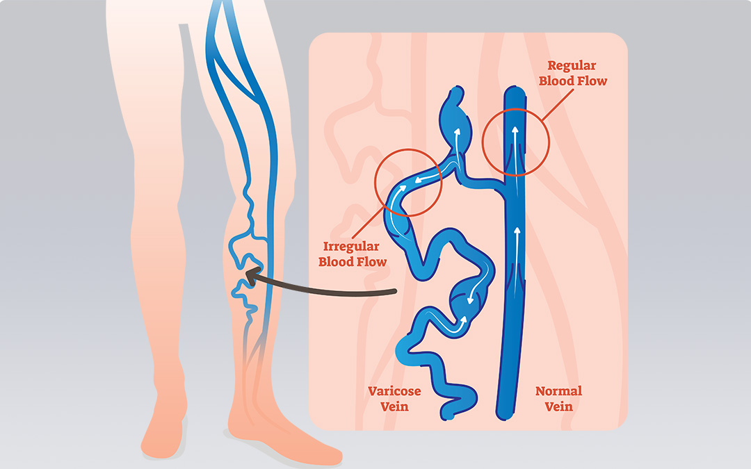
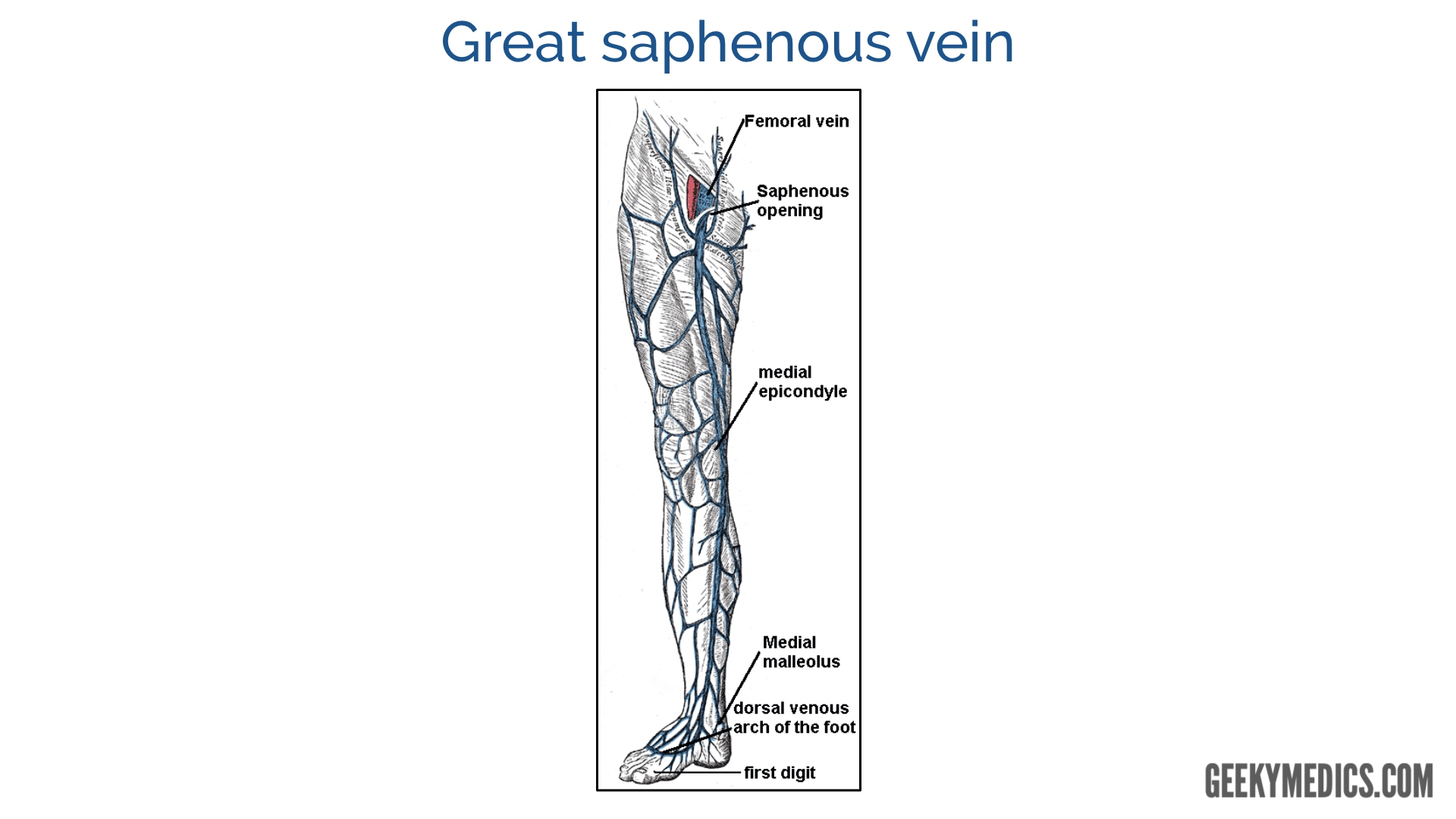
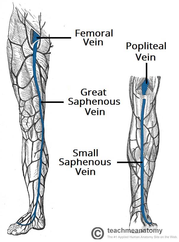
Start studying Leg Veins. Learn vocabulary, terms, and more with flashcards, games, and other study tools. Blood from the dorsal venous arch passes into three major veins in the leg: the small saphenous, great saphenous, and anterior tibial veins. The great saphenous vein ascends through the leg and thigh on the medial side, collecting blood from tissues in these regions. Depending on the extent of valvular insufficiency, blood may pool in the lower parts of the leg and engorge the veins of the leg. Superficial Leg Veins. The two major superficial veins of the leg are the great saphenous and small saphenous veins. The great saphenous vein runs along the front inner (anteromedial) part of the leg and thigh until it joins the femoral vein. The small saphenous travels up the back of the lower leg (posterior aspect). Important veins of the leg include the internal and external iliac veins, femoral vein, saphenous vein, popliteal vein, tibial vein, and the venous arch of the foot. Nerves in the leg send messages...
Great saphenous vein. The great saphenous vein is a large venous blood vessel running near the inside surface of the leg from the ankle to the groin. It arises from the dorsal venous arch at the ... Browse 2,961 veins and arteries diagram stock illustrations and vector graphics available royalty-free, or start a new search to explore more great stock images and vector art. The circulatory or cardiovascular human body system medical illustration. Epifascial, lengthwise tributaries can be distinguished clearly from saphenous veins by ultrasound. Ricci and Caggiati found these veins (superficial accessory veins) in 26 % of legs. They run parallel to the saphenous vein, but in the subcutaneous fatty tissue (Fig. 2.15). This is not duplication of the great saphenous vein. Arteries and nerves of the knee and leg - anterior and posterior views. The popliteal artery is a direct continuation of the femoral artery carrying blood further down the lower limb. In the knee, it gives off the superior and inferior genicular arteries which wrap around this region and supply it with blood.Read about the arterial anastomoses of the lower extremity here.
Thumb Knee Human leg Vein Human anatomy, others, angle, hand ...
Jan 07, 2022 · FIGURE 2.8 Superficial and perforating veins of the leg. nal pudendal veins join each other and the distal GSV to form the confluence of superficial inguinal veins (sapheno-femoral junction) (see Figure 2.9).21 Rarely, the GSV terminates high on the lower abdomen or joins the femoral vein very low and the superficial inguinal veins empty individually into the femoral vein.22 Other occasional tributaries of the GSV in the groin include the posterior and anterior thigh circumflex veins.
Vasculature of the Leg | Texas Heart Institute
Occasionally, veins deep within the legs become enlarged. In such cases, the affected leg may become painful and swell. Any persistent leg pain or swelling warrants medical attention because it may indicate a blood clot — a condition known medically as thrombophlebitis. Bleeding. Occasionally, veins very close to the skin may burst.
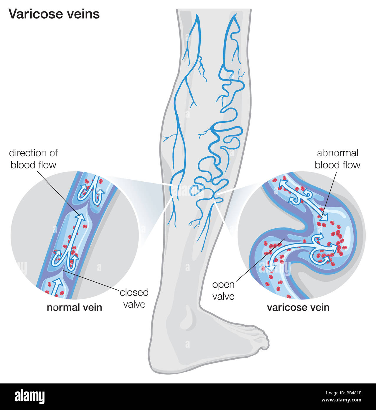
Diagram illustrating varicose veins Stock Photo - Alamy
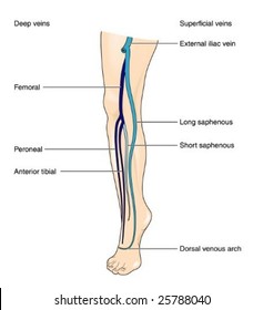
Often, the artery and vein are located within the same vascular sheath - so that the arterial pulsations aid the venous return. The Foot and Leg. The main venous structure of the foot is the dorsal venous arch, which mostly drains into the superficial veins. Some veins from the arch penetrate deep into the leg, forming the anterior tibial vein.

Posterior Arch Vein - Varicose Veins - Flanders Health Blog
3. How to Draw the Artery and Veins Diagram As the cardiovascular system is complex, drawing arteries and veins may seem difficult. If the students follow the step-by-step method, they can make a cardiovascular system connected with arteries, veins, and capillaries. The student can opt for freehand drawing to create a diagram of arteries and veins.
11 Venous Leg Ulcer Illustrations & Clip Art - iStock
Dog leg anatomy. First, you might have a basic idea of the different bones of the forelimb and hindlimb of a dog. Now I will provide you the few information on the other bones of dog leg anatomy with their unique features. The front leg of a dog consists of the clavicle, scapula (arm), radius and ulna (forearm), carpals, metacarpals, and phalanges (forepaw).
varicose veins – redbacteria
Getting older or sitting for long periods of time can also weaken your leg veins and valves. Aneurysm . 14 / 17 . It happens when an artery wall weakens and bulges out like a balloon. Aneurysms ...
Varicose Vein Surgery: Practice Essentials, Anatomy ...
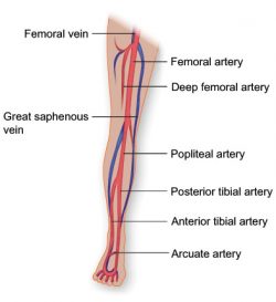
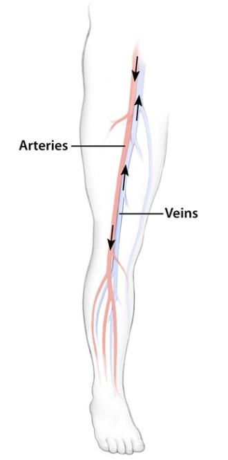
It is the pulmonary artery that brings oxygen-poor blood into your lungs and the pulmonary vein that brings oxygen-rich blood back to your heart. In the diagram showing the vasculature of the leg, the vessels that carry oxygen-rich blood are colored red, and the vessels that carry oxygen-poor blood are colored blue. Email SMS

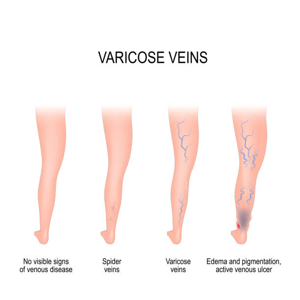
Causes of Varicose Veins | Spider Veins Symptoms | Honolulu
the vein along the posteromedial aspect of the fibula is the peroneal vein. In a US approach, the cortical shadow of the tibia and fibula can be used as a bony landmark. The paired veins are present on both sides of the artery (Fig. 4F). After stretching the patient's leg, the anterior tibial vein can be visualized from an anterolateral

Anterior Leg (Veins) Diagram | Quizlet
Arteries and Veins of the Leg. Create healthcare diagrams like this example called Arteries and Veins of the Leg in minutes with SmartDraw. SmartDraw includes 1000s of professional healthcare and anatomy chart templates that you can modify and make your own. 14/71 EXAMPLES. EDIT THIS EXAMPLE. CLICK TO EDIT THIS EXAMPLE.

Varicose Veins: Causes, Symptoms And Treatment | Netmeds
These veins range in number from 7 to 22, averaging ∼14 per leg, with 52% being direct perforators. 11 The lower perforators tend to be posterior tibial perforators, and the more superior perforators are paratibial. 11 Inferior perforators tend to be short, often only 1 cm in length, whereas toward the middle of the leg they may be 3 to 4 cm ...

Treatment of varicose veins depends on their severity
Veins of the lower limb: Anatomy | Kenhub
Varicose veins - Their cause,symptoms and cure. - HubPages
Varicose Veins | LeMaitre
/GettyImages-87313663-16bdfeaf37d048dbaef06b4f00b269b5.jpg)
Popliteal Vein: Anatomy and Function
Varicose veins by M.Fathy Zaidan

The care of patients with varicose veins and associated ...

Pin on Varicose Veins
lower leg venous anatomy

Schematic representation of leg veins related to wo points ...
A) A 67-year-old woman represented varicose veins as well as ...
261 Varicose Veins Illustrations & Clip Art - iStock

815 Artery Leg Illustrations & Clip Art - iStock
International Vascular Awareness Month | Vein Health Clinic ...

Center for Vein Health - Cardiologists in Gilbert, AZ
Varicose Vein Causes, Symptoms, & Treatments | Calgary

Anatomy of the lower-limb venous system and assessment of ...
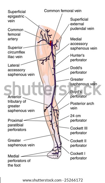
Leg Veins Anterior Labeled Stock Vector (Royalty Free) 25266172
Leg Vein Anatomy | By Vein Specialist in Los Angeles
Veins Leg Labeled Stock Vector (Royalty Free) 25788040
Femoral Vein High Resolution Stock Photography and Images - Alamy

Vein - Wikipedia
Varicose Veins | LeMaitre
Venous Anatomy | Thoracic Key

Send Varicose Veins Packing with this Surprising Change ...

Vein Services | Biltmore Cardiology

Lower Extremity Veins - human anatomy organs
Varicose Vein Examination - OSCE guide | Geeky Medics

Basic anatomy of the venous system of the lower extremities ...
Venous Drainage of the Lower Limb - TeachMeAnatomy


Comments
Post a Comment