40 mitochondria diagram labeled
Mitochondria Label Diagram - cellular respiration, the … 2021-12-20 · Mitochondria Label Diagram. Here are a number of highest rated Mitochondria Label Diagram pictures on internet. We identified it from well-behaved source. Its submitted by government in the best field. mitochondrion the mitochondrion plural mitochondria mitochondrion ultrastructure interactive diagram a mitochondrion has a double membrane printable animal cell diagram - labeled unlabeled and blank printable animal cell diagram to help you learn the organelles in an animal cell in preparation for your test or quiz 5th grade science and biology
Fig: A Labeled Diagram of Chloroplast. Chloroplast Structure Chloroplasts are roughly \(1 - 2\, {\rm{μm}}\) thick and \(5 - 7\, {\rm{μm}}\) in diameter and are seen in all higher plants. In different plants, chloroplasts have different shapes like some plants have filamentous or ovoid or saucer-shaped.

Mitochondria diagram labeled
1. Watch the video on the Mitochondria. 2. As the video plays, (remember: you can pause the video at any time) label everything you see labeled in the video on your own diagram of a mitochondria and a phospholipid bilayer. Mitochondria is the powerhouse of the cell, which produces energy. It is a membrane-bound organelle, present in the cytoplasm of the cell of Eukaryotic organisms which synthesizes energy molecules in the form of ATP, which is used by the cell. Hypothetically mitochondria are believed to have originated as prokaryotic cells like bacteria. Label the organelles in the diagram below. LABEL THE ORGANELLES Word Bank Golgi Apparatus Cell membrane Nucleolus Nucleus Mitochondria Centrioles Lysosome Vesicles Nuclear membrane Flagella Vacuole Ribosomes Endoplasmic reticulum Cytoplasm Microfilament. Animal Cell Worksheet Labeling Worksheets Are Definitely The Spine To Students Learning And Gras In 2021 Animal Cells Worksheet Cells ...
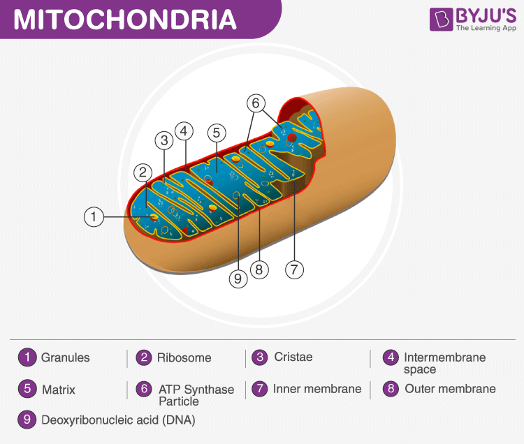
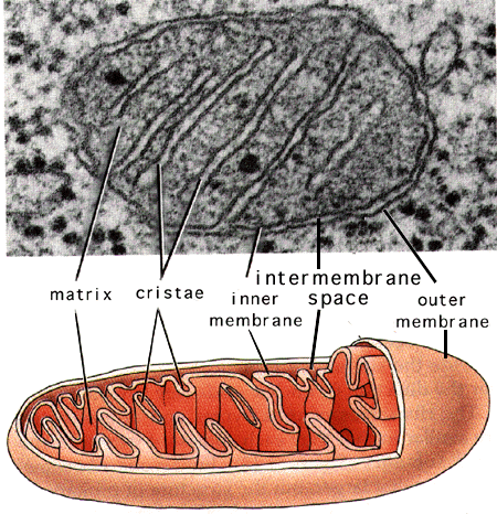
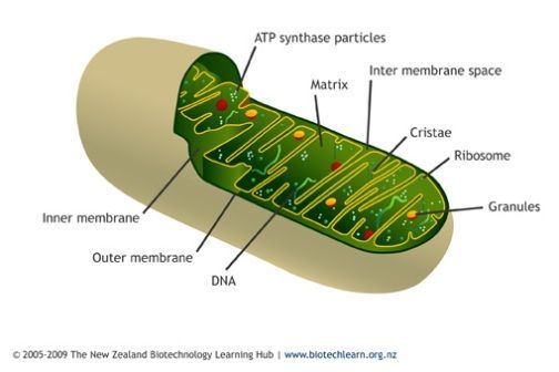
Mitochondria diagram labeled. Diagram Of Mitochondria The diagram below shows the structure and functions of the mitochondria. Structure and Functions Of Mitochondria Matrix It is a viscous or a gel-like fluid containing a mixture of enzymes, ribosomes, inorganic ions, mitochondrial DNA, nucleotide cofactors, and organic molecules. 21.04.2008 · The diagram may be labeled with a version of an mRNA or protein accession (for example, NM_123456.1) different from that listed in the RefSeq section (for example, NM_123456.2). This will result if the sequence has been changed in any way, such as extending the 5' or 3' ends, or removing mismatches between the cDNA sequence and the reference … Human Cell Diagram, Parts, Pictures, Structure and Functions The cell is the basic functional in a human meaning that it is a self-contained and fully operational living entity. Humans are multicellular organisms with various different types of cells that work together to sustain life. This BiologyWise article the structure and function of mitochondria with the help of a labeled diagram. The study of cell organelles and their functions covers individual responsibilities of each subunit of a cell - each organelle is a structure within a cell that has a specific function.
How Many Membranes Are Present In Mitochondria Labeled. Saturday, May 15th 2021. ... that the human physique is very elaborate and a technique I learned to are aware of it is by means of the way of human anatomy diagrams. Many folks have didn't comprehend the numerous details, as students, or patients whilst your medical professional has ... Animal cell diagram mitochondria.Animal cells Almost all animals and plants are made up of cells. A bacteria diagram clearly helps us to learn extra about this single cell organisms that have neither membrane-bounded nucleolus or organelles like mitochondria and. Centrioles are about 500nm long and 200nm in width that are found close to the nucleus and helps in cell division. Mitochondria occupy a substantial portion of the cytoplasmic volume of eucaryotic cells, and they have been essential for the evolution of complex animals. Without mitochondria, present-day animal cells would be dependent on anaerobic glycolysis for all of their ATP. When glucose is converted to pyruvate by glycolysis, only a very small fraction of the total free energy potentially available ... Mitochondrion ultrastructure (interactive diagram) a mitochondrion has a double membrane; The stages of cellular respiration include glycolysis, pyruvate oxidation, the citric acid or krebs cycle, and oxidative phosphorylation. This series of reactions produces 36 molecules of atp! Please update your bookmarks accordingly.
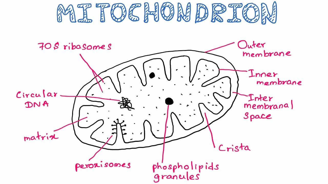
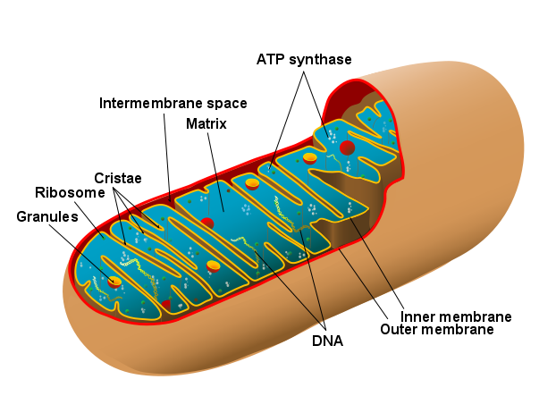
04.03.2021 · A bacteria diagram clearly helps us to learn extra about this single cell organisms that have neither membrane-bounded nucleolus or organelles like mitochondria and chloroplasts. They are obviously a cause of diseases to men and women and animals, yet their helpful facets cannot be ignored. For instance, sure bacteria like actinomycetes produce … 17.06.2021 · Mitochondria: 1. Double-membrane bound, a powerhouse of the cell. 2. This is an autonomous organelle. 3. The outer membrane is smooth and the inner membrane is highly folded. 4. The inner membrane forms cristae on which oxysomes are present. 5. The matrix contains 70S ribosomes, few RNA molecules and circular, naked, dsDNA. 6. They are involved ... Mitochondria are usually rod-shaped or sausage-shaped, and are double-membraned structures made of an outer membrane which surrounds the organelle, and an inner membrane which contains many finger-like folds called Cristae. Diagram of Mitochondria Mitochondria have their own DNA and ribosomes, different from the rest of the cell. 16.08.2021 · Cancer isn’t usually top of mind for young women, but this type -- caused by the human papillomavirus (HPV) -- is a serious threat. Each …
Bio Geo Nerd: January 2013
Functions of Mitochondria. The most important function of the mitochondria is to produce energy. The simpler molecules of nutrition are sent to the mitochondria to be processed and to produce charged molecules. These charged molecules combine with oxygen and produce ATP molecules. This process is known as oxidative phosphorylation.
how to draw the mitochondria - YouTube
Animal Cell Diagram Mitochondria Labeled Tuesday, April 27th 2021. | Diagram Animal Cell Diagram Mitochondria. Animal cells have a basic structure. The mitochondrion (plural mitochondria) is a membrane-bound organelle found in the cytoplasm of eukaryotic cells.
What are Bacteria - iFink!
A well-labelled diagram of mitochondria is given below for your better understanding of the structure. Labelled Diagram of a Mitochondrion Image will be updates soon Characteristics of the Mitochondrial DNA/ Genome: The mitochondrial DNA is circular and is made up of 16,569 DNA base pairs.
Chemical Composition of Mitochondria with Definition and ...
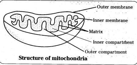
Mitochondria are mobile, plastic organelles that have a double-membrane structure. It ranges from 0.5 to 1.0 micrometer in diameter. It has four distinct domains: the outer membrane, the inner membrane, the intermembrane space, and the matrix. The organelle is enclosed by two membranes—a smooth outer membrane and a markedly folded or tubular ...

Describe the structure of mitochondria with the help of a ...
Mitochondria are energy-producing organelles found in most living cells. They use carbohydrates such as glucose in chemical reactions based on an electron transport chain and the citric acid cycle. The final products of these reactions are water and ATP, an energy-storage molecule.

Mitochondria Functions - Biology Wise
Mitochondria diagram explaining the structure of mitochondria Structure of Mitochondria The mitochondrion is a double-membraned, rod-shaped structure found in both plant and animal cell. Its size ranges from 0.5 to 1.0 micrometre in diameter. The structure comprises an outer membrane, an inner membrane, and a gel-like material called the matrix.

Mitochondria Structure Pencil Diagram - Diagram Media
Start studying Mitochondria Labeling. Learn vocabulary, terms, and more with flashcards, games, and other study tools.

Places To Be , Hamburg
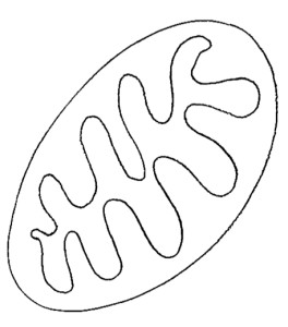
Mitochondria are cell organelles whose overall shape resembles rounded rods and is often drawn in 2D as an oval-shape. Mitochondria have a double-membrane structure and contain many substructures including enzymes, ribosomes and mitochondrial DNA (mtDNA). Structure of mitochondria is useful for A-Level biology.

The Trenches of Discovery: October 2013
Summary. Description. Animal mitochondrion diagram unlabelled.svg. English: A diagram showing a mitochondrion of the eukaryotic cell. Mitochondria are organelles surrounded by membranes, distributed in the cytosol of most eukaryotic cells. Its main function is the conversion of potential energy of pyruvate molecules into ATP.

#94 Structure and function of the mitochondrion | Biology ...
A Labeled Diagram of the Animal Cell and its Organelles. There are two types of cells - Prokaryotic and Eucaryotic. Eukaryotic cells are larger, more complex, and have evolved more recently than prokaryotes. Where, prokaryotes are just bacteria and archaea, eukaryotes are literally everything else. From amoebae to earthworms to mushrooms, grass ...

Diagram Mitochondria Sketch - Aflam-Neeeak
Download mitochondria-diagram-labeled : Filetype: Size: EPS/Vector format Available for Purchase: PNG with Transparent Background (Members) GIF with transparent Background (Members) Large JPG (Members) Medium JPG (Members) Free Download: 87 Kb: To Purchase Clipart as Vector File contact support@classroomclipart.com
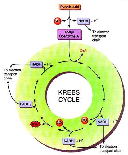
Aerobic Respiration, Part 2: Oxidation of Pyruvate and The ...
labeled mitochondria showing the looping of the reticulum throughout its length. transitioning of mitochondria between punctate and reticulum states through alternating fission and fusion is now known to be critical to maintaining mitochondrial quality control. Fission allows separation of

Science Doing: Mitochondrion Cell Organelle: A Symbiotic ...
Mitochondria are double membrane-bound cell organelles responsible for the supply and storage of energy for the cell. The oxidation of various substrates in the cell to release energy in the form of ATP (Adenosine Triphosphate) is the primary purpose of mitochondria. ... Structure, Parts, Functions, Labeled Diagram, Worksheet; Animal Cell ...

The structure of a mitochondrion | Download Scientific Diagram
This will also help you to draw the structure and diagram of mitochondria. 1. Mitochondria are commonly called the "Power house" of the cell. 2. Benda (1897) was the first to coin the term mitochondrion. 3. Usually, mitochondria are 0.5 to 1 n in diameter and 3-6n in length.

Labelled Diagram Of A Human Cell Bone Cell Labeled Diagram ...
Label the organelles in the diagram below. LABEL THE ORGANELLES Word Bank Golgi Apparatus Cell membrane Nucleolus Nucleus Mitochondria Centrioles Lysosome Vesicles Nuclear membrane Flagella Vacuole Ribosomes Endoplasmic reticulum Cytoplasm Microfilament. Animal Cell Worksheet Labeling Worksheets Are Definitely The Spine To Students Learning And Gras In 2021 Animal Cells Worksheet Cells ...

Closeup of skeleton foot model
Mitochondria is the powerhouse of the cell, which produces energy. It is a membrane-bound organelle, present in the cytoplasm of the cell of Eukaryotic organisms which synthesizes energy molecules in the form of ATP, which is used by the cell. Hypothetically mitochondria are believed to have originated as prokaryotic cells like bacteria.

Mitochondria Anatomy
1. Watch the video on the Mitochondria. 2. As the video plays, (remember: you can pause the video at any time) label everything you see labeled in the video on your own diagram of a mitochondria and a phospholipid bilayer.
Labeled Hand Drawn Mitochondria Diagram - Diagram Media
Draw a diagram of human sperm. Label only those parts ...

santhoshproject

Structure and function of mitochondrial membrane protein ...

Mitochondrial respiration. 24 Q CO2 , carbon dioxide ...

Cell Biology: mitochondrion

The Nucleus of the Cell and Related Organelles
A Labelled Diagram Of Mitochondria with Detailed Explanation

Assignment 5, page 3

Mitochondria - CoolaBoo - Education Site

Diagram Mitochondria Sketch - Aflam-Neeeak

Mitochondria – An overview of structure and function
Mitochondria - microbewiki

Chloroplasts & Mitochondria *Compare and contrast the ...

Modified Q-cycle and ROS generation in complex III. The ...
More about mitochondria — Science Learning Hub

Kevin Ahern's Biochemistry (BB 451/551) at Oregon State ...
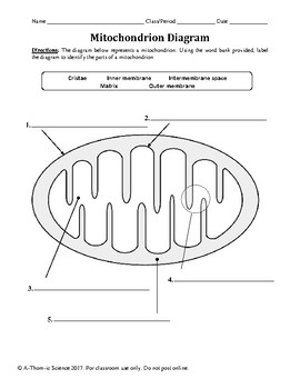
Mitochondrion & Cellular Respiration Diagram Worksheet by ...

Mitochondria, Neuropathy, HIV and Fluoroquinolones ...
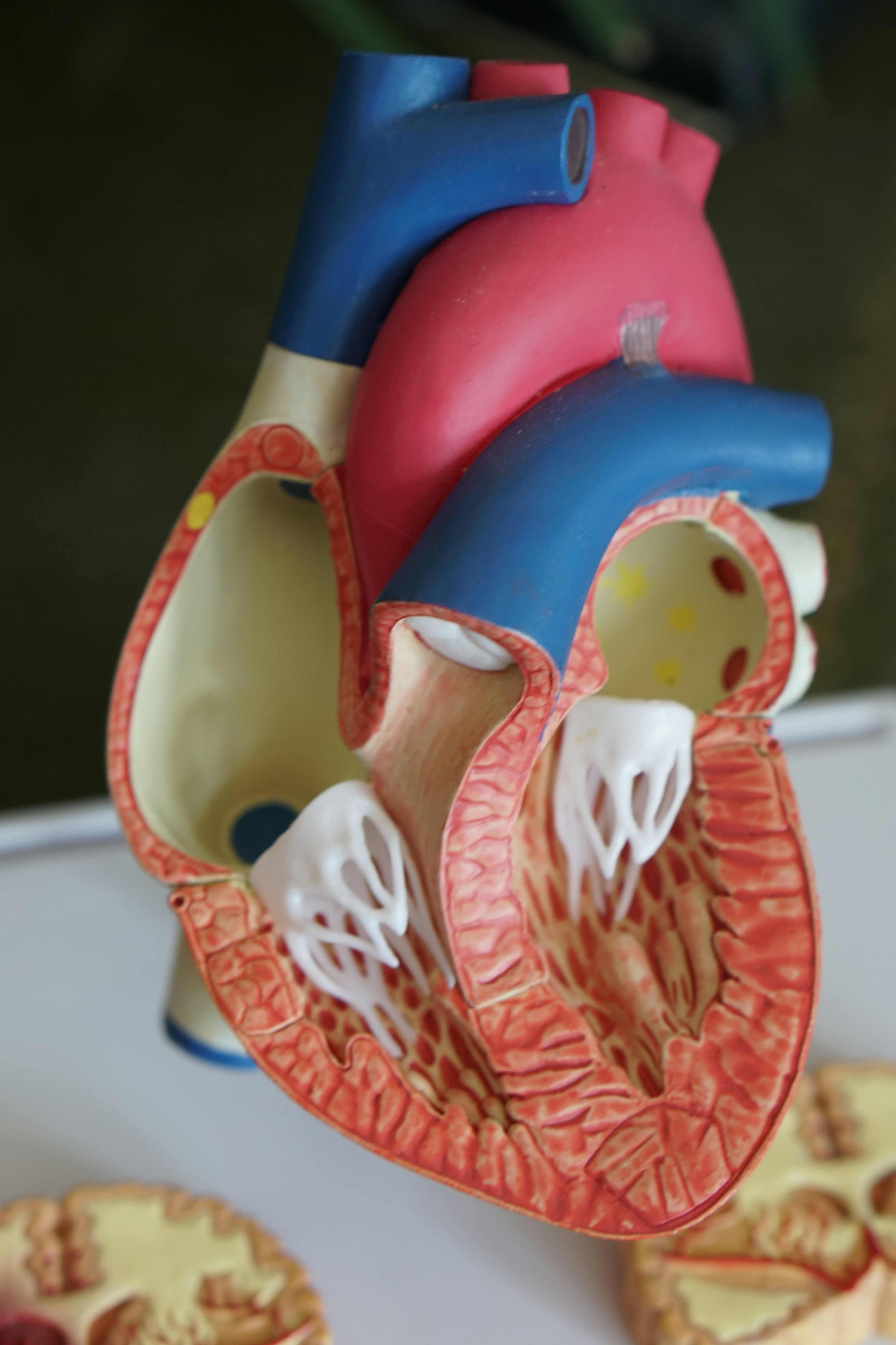
Open heart model

Cellular Respiration Mitochondria Diagram Labeled ...

Mitochondrial structure adapted by Freitas [41 ...

Mitochondria - CELLULAR RESPIRATION


Comments
Post a Comment