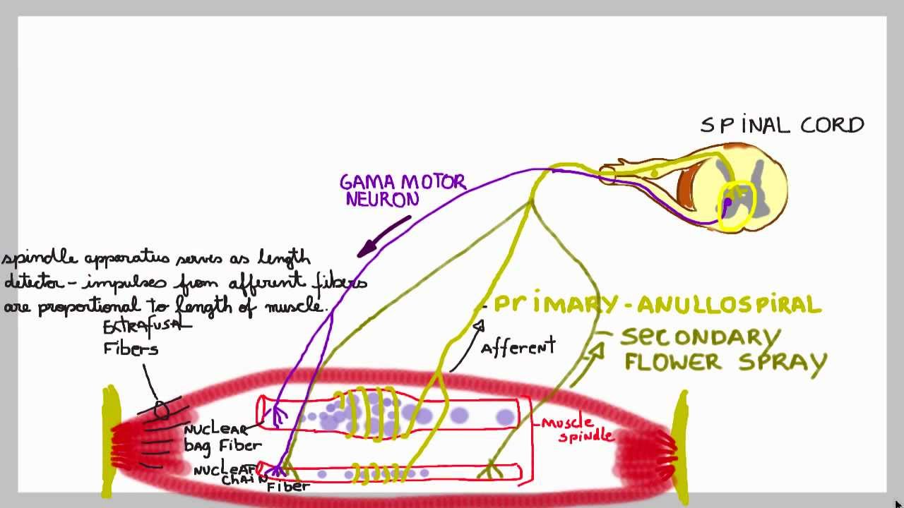
38 muscle spindle diagram
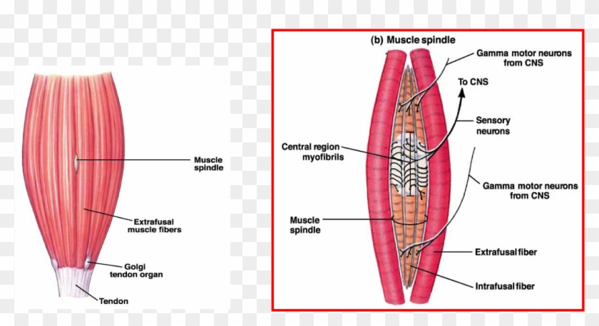
Muscle spindles are sensitive to changes in velocity and are innervated by type 1a nerve fibres. These afferent nerve fibres conducting the impulse directly to the spinal cord, where they are immediately conducted via interneurons to alpha motor neurons, which stimulate muscle contraction. organization of the muscle spindle is shown in Figure 54-2. Each spindle is 3 to 10 millimeters long. It is built around 3 to 12 very small intrafusal muscle fibers that are pointed at their ends and attached to the glycoca-lyx of the surrounding large extrafusal skeletal muscle fibers. Each intrafusal muscle fiber is a very small skeletal muscle fiber.
Muscle spindles are ubiquitous encapsulated mechanoreceptors found in most mammalian muscles. There are two types of endings, primary and secondary, and both ...
Muscle spindle diagram
(A) Diagram of muscle spindle, the sensory receptor that initiates the stretch reflex. (B) Stretching a muscle spindle leads to increased activity in Ia afferents and an increase in the activity of α motor neurons that innervate the same muscle. September 15, 2018 - This article describes three simple activities we presented at the 2017 FUN Faculty Workshop at Dominican University that demonstrate how proprioceptive information contributes to our mental image of physical self, and how artificially altering this information ... Anatomy and Physiology. Anatomy and Physiology questions and answers. Om Label on the diagram: 1. Muscle spindle 2. Sensory neuron 3. Soma of sensory neuron 4. Dorsal root 5. Dorsal root ganglion 6.
Muscle spindle diagram. October 8, 2014 - Muscle spindles are commonly considered as stretch receptors encoding movement, but the functional consequence of their efferent control has remained unclear. The “α–γ coactivation” hypothesis states that activity in a muscle is positively related to the output of its spindle afferents. Describe a diagram showing intrafusal and extrafusal muscles fibres with innervation Contraction of the muscle spindle - contractile proteins at polar ends and none in the equatorial region, therefore the polar ends can contract but the fluid filled equatorial region cannot Image 2: Mammalian muscle spindle showing typical position in a muscle (left), neuronal connections in spinal cord (middle) and expanded schematic (right). The stretch reflex is designed as a protective mechanism, to prevent strain and tear injuries to the muscles and tendons.When the muscle spindle is excited an impulse is immediately received to contract the muscle, thereby protecting it from being pulled forcefully or stretched beyond a normal range of motion.
The information on the diagram opposite led to the following conclusions regarding the function of the muscle spindle: The muscle spindle is a Length Receptor that senses changes in the length of the muscle. In particular stretch causes an increased rate of firing of action potentials (A) The muscle spindle is unloaded (i.e. shortens and has a reduced tension) during contraction of the extrafusal fibres (B), November 30, 2020 - Other articles where muscle spindle is discussed: human nervous system: Muscle spindles: The familiar knee-jerk reflex, tested routinely by physicians, is a spinal reflex in which a brief, rapid tap on the knee excites muscle spindle afferent neurons, which then excite the motor neurons of ... Q. 1 Muscle spindle detects: Muscle spindle and Golgi tendon organ both receptors are important component of intrinsic muscle control. Muscle spindle acts through muscle stretch reflex. Whenever a muscle is stretched suddenly, excitation of the spindles cause reflex contraction of the large skeletal muscle fibers of stretched muscle thus ... Muscle spindles are stretch receptors within the body of a skeletal muscle that primarily detect changes in the length of the muscle. They convey length information to the central nervous system via afferent nerve fibers.This information can be processed by the brain as proprioception.The responses of muscle spindles to changes in length also play an important role in regulating the ...
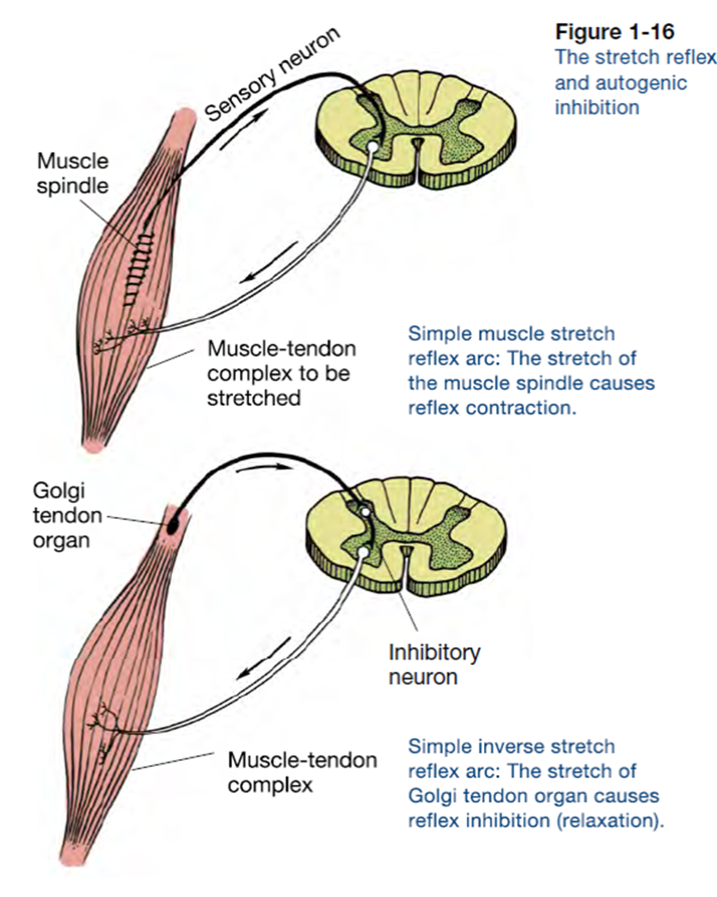
with the 'stretch reflex' (Diagram 1): l Muscle spindles sensitive to change in length and speed of change in muscle fibres. l Golgi tendon organs that detect prolonged change in tension. Stretching a muscle causes an increase in the impulses transmitted from the muscle spindle to the posterior horn cell (PHC) of the spinal cord. Fig. 2 Examples of muscle-spindle pr imary ending s respondin g to trap- ezoidal ( a , c ) and sinusoida l ( b , d ) stretc hes applied to th e tendon of the muscle (per oneus tertiu s of cat). Download scientific diagram | Staining patterns of muscle spindle in the B region. Serial transverse sections stained for mATPase at pH 4.6 ( A ), and stained with antibodies against MyHCsto ( B ... The next shot demonstrates that the muscle spindle and the extra fuse all muscle are innervated by different motor now or five verse and that the gamma fuse in motor fibers have a higher threshold to stimulation than the alpha fibers the spindle is exposed in the muscle but not isolated repetitive stimulation of Alpha fibers unloads the spindle ...
Golgi Tendon Organs and Muscle Spindles Explained | ACE
Fig. 1.Simplified diagram of the central region of a muscle spindle in the cat, showing a single “bag” intrafusal muscle fiber and a single “chain” intrafusal muscle fiber. Typically, muscle spindles in the cat contain 2 or 3 bag fibers, 4–6 chain fibers, and a complex motor innervation.

Santa Monica Presidents Day 2018, when you love America so much you take to the streets and sing to your people.
Muscle spindles can be defined as small, spindle-shaped sensory receptors located in skeletal muscle tissue (Fig. 6.11), and they run parallel to the main muscle fibers (extrafusal fibers). A muscle spindle consists of several differentiated muscle fibers (intrafusal fibers) that are enclosed in a spindle-shaped connective tissue sac.

Declined push-up
Official Ninja Nerd Website: https://ninjanerd.orgNinja Nerds!In this lecture Professor Zach Murphy will present on the spinal cord stretch reflex, in additi

Muscle Spindles / Golgi Tendon Organs - Neurology ...
January 7, 2021 - Almost every muscle contains muscle spindles. These delicate sensory receptors inform the central nervous system (CNS) about changes in the length of individual muscles and the speed of stretching. With this information, the CNS computes the position and movement of our extremities in space, ...

Neurones.co.uk
**Diagram a stretch reflex with alphagamma coactivation in the muscle spindle. *** it would be nice to have a better explanation of this Excitation of gamma motor neurons and alpha motor neurons at the same time is a process known as alpha-gamma coactivation.

Closeup of skeleton foot model
Label on the diagram: 1. Muscle spindle 2. Sensory neuron 3. Soma of sensory neuron 4. Dorsal root 5. Dorsal root ganglion 6. Dorsal horn 7. Ventral horn 8. Motor neuron to quad 9.

Image from page 440 of "Principles of modern biology" (1964)
The muscle spindle is a proprioceptor. a sense organ that receives information from muscle, that senses STRETCH and the SPEED of the stretch.

View of a tree on the bluff overlooking the Pacific Ocean
The meaning of MUSCLE SPINDLE is a sensory end organ in a muscle that is sensitive to stretch in the muscle, consists of small striated muscle fibers richly supplied with nerve fibers, and is enclosed in a connective tissue sheath —called also stretch receptor.

Muscle spindle (3-4mm), Extrafusal intrafusal muscle ...
Muscle spindles (important for muscle length and velocity and to a lesser extent, monitoring static length) Golgi tendon organs (detects muscle force) Joint afferents (most sensitive to position at extreme joint angles) Tactile (pressure receptors in muscle and overlying skin, sense flexion or extension of finger) 1st Required QUIZ. UNIT 4.

Baby gull calling to a parent for food
Examples of muscle-spindle primary endings responding to trapezoidal (a, c) and sinusoidal (b, d) stretches applied to the tendon of the muscle (peroneus tertius of cat).a, b The reproducibility of the responses when five separate presentations of the stimuli are given to the same primary ending. The responses are superimposed and each response is indicated by different coloured symbols.

Muscle spindle Illustrations
Muscle spindles are small sensory organs with an elongated shape, involved in proprioception. Image 2: Mammalian muscle spindle showing typical position in a muscle (left), neuronal connections in spinal cord (middle) and expanded schematic (right). The spindle is a stretch receptor with its own motor supply consisting of several intrafusal muscle fibres.

Muscle Spindle Apparatus - Human Physiology - 78 Steps ...
In this video, we review GTOs and discuss the functions of muscle spindles in providing proprioceptive information about muscle length and ...

Muscle spindle Illustrations
The pathway starts when the muscle spindle is stretched (caused by the tap stimulus in the knee jerk reflex). The muscle spindles are responsible for detecting the length of the muscles fibres. When a stretch is detected it causes action potentials to be fired by Ia afferent fibres. These then synapse within the spinal cord with α-motoneurones ...

Simplified diagram of the central region of the muscle ...
muscle contraction (both extra- and intra-fusal fibres) decreased firing of spindle afferent (negative feedback) co-activation of both the alpha and gamma motor neurons (step 3) prevents the spindle afferents from becoming completely silent during step 5 (i.e., the gap in firing shown in the diagram doesn't happen in real life!)

Muscle spindle has both sensory and motor function - YouTube
May 2, 2017 - Learn about the two most basic underlying structural components of the body, Golgi tendon organs and muscle spindles, and how they work together.
Golgi Apparatus Simple Diagram - Muscle Spindle Parts, HD ...
Proprioception is the sense of the body’s position in space based on specialized receptors that reside in the muscles and tendons. The muscle spindle signals the length of a muscle and changes in the length of a muscle. The Golgi tendon organ signals the amount of force being applied to a muscle.
Terms
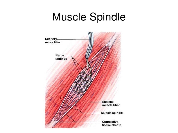
Muscle spindles are sensory receptors that are located in muscle. Their job is to detect changes in muscle length and the speed of change in muscle length. Below is an image of a muscle spindle. When muscles lengthen, the spindles are stretched. This stretch activates the muscle spindle which in turn sends an impulse to the spinal cord.

Image from page 687 of "A manual of diseases of the nervous system" (1907)
A: sensory innervation of skeletal muscles. The size of the receptors to the muscle exaggerated. Note that the muscle spindle is attached via connecgtive tissue fibers to the tendons. Thus, muscle spindle wherever the whole muscle is stretched. B: Schematic representation of the two kinds of intrafusal muscle fibers and their innervation (Brodal)

Study Notes
muscle contraction (both extra- and intra-fusal fibres) decreased firing of spindle afferent ( negative feedback ) co-activation of both the alpha and gamma motor neurons (step 3) prevents the spindle afferents from becoming completely silent during step 5 (i.e., the gap in firing shown in the diagram doesn't happen in real life!)

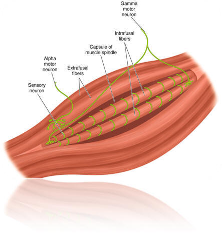
Detailed innervation of a muscle spindle. Schematic of an ...
The Muscle Spindle is a proprioceptive receptor composed of intrafusal fibers that are located within the extrafusal fibers of skeletal muscle. The intrafusal fibers of the muscle spindle run parallel to the extrafusal fibers of the muscle, attaching to them along short segments.

| Diagrammatic representation of muscle spindle ...
Download scientific diagram | | Diagrammatic representation of muscle spindle. from publication: Spasticity Mechanisms – for the Clinician | Spasticity, a classical clinical manifestation of an upper motor neuron lesion, has been traditionally and physiologically defined as a velocity dependent ...

Image from page 58 of "Elements of biology, with special reference to their rôle in the lives of animals" (1933)
(A) Diagram of a biologic muscle spindle and a model of spindle structure. A muscle spindle consists of three types of intrafusal fibers that receive fusimotor inputs (gamma static and dynamic) while giving rise to primary (Ia) and secondary (II) afferents. (B) Model of spindle output during 6-mm whole-muscle ramp stretches. Primary afferent ...

Muscle Spindles / Golgi Tendon Organs - Neurology ...
January 7, 2021 - Almost every muscle contains muscle spindles. These delicate sensory receptors inform the central nervous system (CNS) about changes in the length of individual muscles and the speed of stretching. With this information, the CNS computes the position and movement of our extremities in space, ...

Right Side Chest pose Over 60 Bodybuilding and Fitness
Anatomy and Physiology. Anatomy and Physiology questions and answers. Om Label on the diagram: 1. Muscle spindle 2. Sensory neuron 3. Soma of sensory neuron 4. Dorsal root 5. Dorsal root ganglion 6.

Understanding the Stretch Reflex (or Myotatic Reflex)
September 15, 2018 - This article describes three simple activities we presented at the 2017 FUN Faculty Workshop at Dominican University that demonstrate how proprioceptive information contributes to our mental image of physical self, and how artificially altering this information ...

PORTRAITS INSTAGRAM - @LGNWVRPRTRTS EDITORIAL INSTAGRAM - @LGNWVRPHTO PERSONAL INSTAGRAM - @LGNWVR
(A) Diagram of muscle spindle, the sensory receptor that initiates the stretch reflex. (B) Stretching a muscle spindle leads to increased activity in Ia afferents and an increase in the activity of α motor neurons that innervate the same muscle.
L5 Muscle Structure

Figure 1 from Muscle Spindle and Comparison of Fish Muscle ...

POOL DAY
Five hours of makeup and paint to achieve the human anatomy photoshoot. Thank you Steph and Shay. See more and official credit on @jawfox.photography.

Fit man

Three positions of the muscle spindle. (1) Relaxed muscle ...

Single leg push-up lowered
Muscle spindles: basic mechanism of these stretch sensors ...

Image from page 628 of "Anatomischer Anzeiger" (1897)

Handsome young man with killer abs

Muscle Spindles

san diego


Comments
Post a Comment