38 dog eye diagram
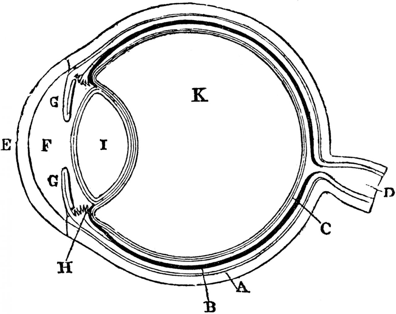
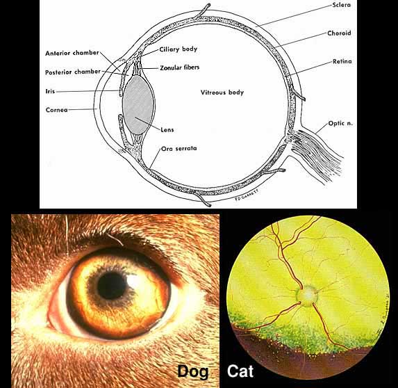
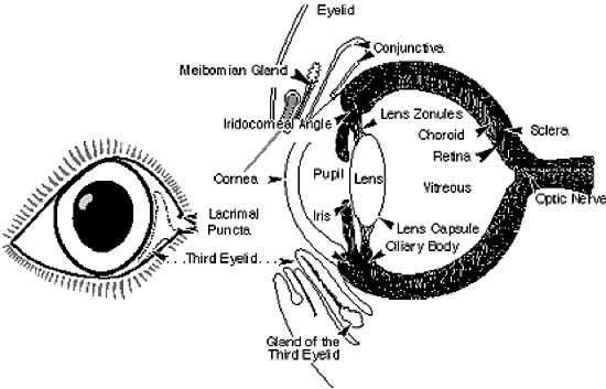
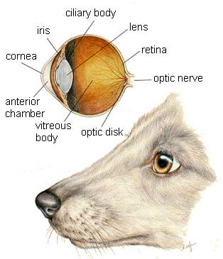
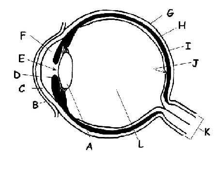
The following diagram and paragraphs explain the skeletal anatomy of a dog. One extremely important part of a dog's skeletal anatomy is the skull. It is a long bone structure that encases the brain, and contains a cavity called the orbit, where the eye is located. Dog Eye Anatomy. " Dog Eye Anatomy is a reflection of the eye as a complicated organ, containing many parts. These functional parts, i.e. sclera, conjunctiva, cornea, iris, pupil, lens, retina, lacrimal gland etc. are contained in a bony socket called a "orbit". Along with these dog eye parts, muscles, blood vessels, nerves and tear drains ...
Jul 26, 2014 - Learn more about pet care options at VETSS, a full-service veterinary hospital and 7 day per week ... Dog canine eye anatomy illustration.

Dog eye diagram
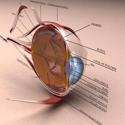
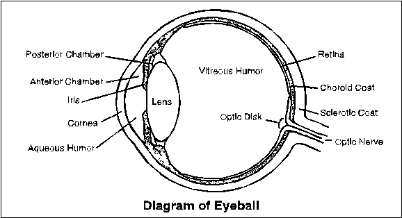
Anatomy of the eye. The bony cavity or socket that contains the eyeball is called the orbit. The orbit is a structure that is formed by several bones. The orbit also contains muscles, nerves, blood vessels, and the structures that produce and drain tears. The white of the eye is called the sclera. This is the relatively tough outer layer of the ... A bull's eye diagram is a strategic template designed in the shape of a bull's eye. It helps teams work out where their real priorities lie. Each concentric circle radiating out from the center corresponds to a different level of importance. The inner-circle should hold only the absolute top-priority tasks, all the way to the outermost ... Dogs therefore have two sets of teeth, baby teeth (28), which will eventually fall out, and adult teeth (42). Different types of dog teeth. As you can see in the diagram above, there are different types of dog teeth. These different types of dog teeth include Incisors, Canine, Premolars, and Molars. How many teeth do dogs have on top
Dog eye diagram. dog eye diagram eye diagram illustration. old engraved illustration of eye infections and diseases - eye diagram stock pictures, royalty-free photos & images. various medical operations engraving antique illustration, published 1851 - eye diagram stock illustrations. Arterial Blood Vessels of the Orbit. Page 5. General Anatomy. Dog. Blood Supply. Horizontal section. Long Posterior Ciliary a. Blood enters the globe. Short ...77 pages Anatomy of the dog - Illustrated atlas This modules of vet-Anatomy provides a basic foundation in animal anatomy for students of veterinary medicine. This veterinary anatomical atlas includes selected labeling structures to help student to understand and discover animal anatomy (skeleton, bones, muscles, joints, viscera, respiratory system ... In a Dog's Eye. The visual system is an important sense available to the canine. ... Distichiasis is a condition in which small hair structures abnormally grow on the inner surface of the eyelids ( see diagram ). Both upper and lower lids may be involved. The abnormal hairs growing on the inner surface of the lids cause irritation to the cornea.
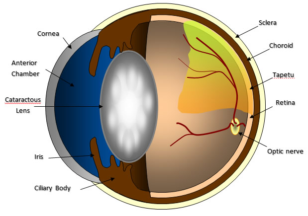
Mammal Prairie Dog, Black-tailed Orange Mammal Cat, Domestic Red or Orange With orange, brown or blue eyes Mammal Fox, Red Red or Orange Medium Mammal Hares Red or Orange Mammal Rabbits Red or Orange Mammal Rat, Norway/Black Red or Orange Bird Nighthawks Red Pinkish Bird Owl, Barn Red Dull Bird Owl, Barred Red Strong Anatomy of the cat`s or dog`s eye. Royalty-Free Vector. Download preview. Anatomy of the cat`s or dog`s eye. Vertical section of the eye and eyelids. Third eyelid and Tapetum lucidum. Schematic diagram. detailed illustration. dog eye anatomy, cat anatomy, How to draw dog eyes step by step Step 1: Mark the page with two directional lines that divides the dog's face in half. This helps with the symmetry of the face and lines up the eyes at one level. Pay attention to the fact that the face is round, not flat — the line marking the general position of the eyes is a curve. Oct 29, 2021 — Dog Eye Anatomy · Sclera: Tough, fibrous layer that's often referred to as the “white” of the eye · Cornea: Thin, clear layer at the front of the ...
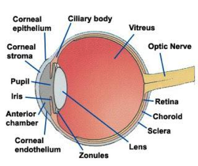
They contain a tarsal plate (for support), muscle, glands and cilia (eyelashes). The outer skin is thin when compared to the rest of the body. Neither the dog ...May 31, 2019 · Uploaded by Dechra Pharmaceutical A simple eye injury is one that penetrates or perforates the cornea or sclera of the eye - notice in the diagram above that these are injuries to the outer part of the eye. An example of a penetration would be a splinter piercing, whereas a perforation would be more like a scratch that goes across this part of the eye. The eye is a paired organ, the organ of vision. The eye is made up of various components, which enable it to receive light stimuli from the environment, and deliver this stimuli to the brain in the form of an electrical signal. Vision involves all components of the eye. Structure. The eye is contained within the bony orbit of the head. Anatomy of the eye. The bony cavity or socket that contains the eyeball is called the ...
Dragonaters: On the road with Motorsports Psychology and ...
Dog anatomy comprises the anatomical studies of the visible parts of the body of a domestic dog.Details of structures vary tremendously from breed to breed, more than in any other animal species, wild or domesticated, as dogs are highly variable in height and weight. The smallest known adult dog was a Yorkshire Terrier that stood only 6.3 cm (2.5 in) at the shoulder, 9.5 cm (3.7 in) in length ...

Dog day out
Labeled anatomy of the head and skull of the dog on CT imaging (bones of cranium, brain, face, paranasal sinus, muscles of head) This module of vet-Anatomy presents an atlas of the anatomy of the head of the dog on a CT. Images are available in 3 different planes (transverse, sagittal and dorsal), with two kind of contrast (bone and soft tissues).
Doggy without a home
Differences Similarities. -less cone in the retina -Eyes located on the front of the skull. -Can only see small amount of colour -Same general structure. -Cant see stationery objects clearly. -Can see things clearer in motion. -Dogs can see in the dark. In the image above the top is what the human eye sees and the bottom is what the a dog sees.

Eye | anatomy | Britannica.com
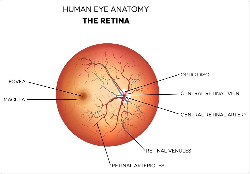
Vision is a complex phenomenon in which light emanating from objects in the environment is captured by the eye and focused onto the retinal photoreceptors (Figures 1-1 and 1-2).Electrical signals originating from these cells pass through a number of cell types in the retina and throughout the central nervous system (CNS) before arriving at the visual cortex, where the sensation of vision occurs.
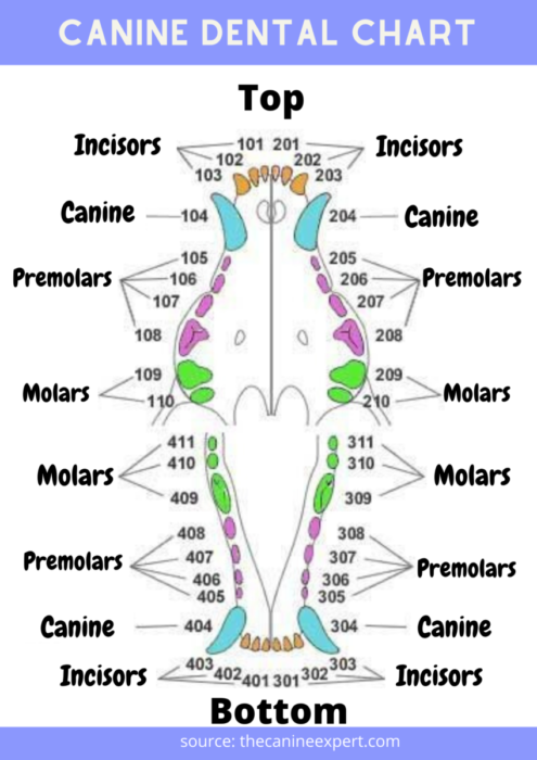
Canine Dental Chart: Dog Teeth Diagram - KittyExpert.com
Browse 4,450 eye diagram stock photos and images available, or search for human eye diagram or eye diagram vector to find more great stock photos and pictures. Vintage anatomical color illustration of the muscles and bones of the face showing the eyeball and the circumorbital area. Diagram Of A Retinal Detachment.
Unlabeled Eye Diagram - ClipArt Best
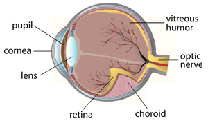
The dog's eye is pretty much a garden-variety mammalian eye, with some notable adaptations that have evolved over the millennia. It is a globe with two fluid-filled chambers (anterior and posterior). The chambers are separated by the lens, the structure that helps focus light beams onto the rear part of the eye, the retina.

Animals with ocular diseases. (A) Eye, dog. Red irregular ...
Dog's view. Visual acuity. Visual acuity is a measure of the spatial resolution of the visual system. It is often measured in cycles per degree (CPD), which measures how much an eye can differentiate one object from another in terms of visual angles. The maximum visual acuity of the human eye is around 50 CPD [7] and 60 CPD [8].
Fluffy cockapoo having the time of his life at the park
An eye diagram is used in electrical engineering to get a good idea of signal quality in the digital domain. To generate a waveform analogous to an eye diagram, we can apply infinite persistence to various analog signals a well as to quasi-digital signals such as square wave and pulse as synthesized by an arbitrary frequency generator (AFG).
Carnivore Anatomy Lab 24 Introduction
Sep 9, 2016 - Anatomy Of A Dogs Eye ed79a0172cea6d61ed91bdca6a2a38a4…

Frog Sculpture, Dunston, Gateshead, Tyne & Wear, England.
Close-up face of Cute pug puppy dog sleeping rest open eye by chin. Border collie dog with different eye color. Picture showing a border collie dog with different eye color - blue/white and brown. Hungry funny puppy dog licking its nose with tongue out and winking one eye closed. Isolated on blue colored background.

dog eye anatomy - Αναζήτηση Google | Rectus muscle, Eye ...
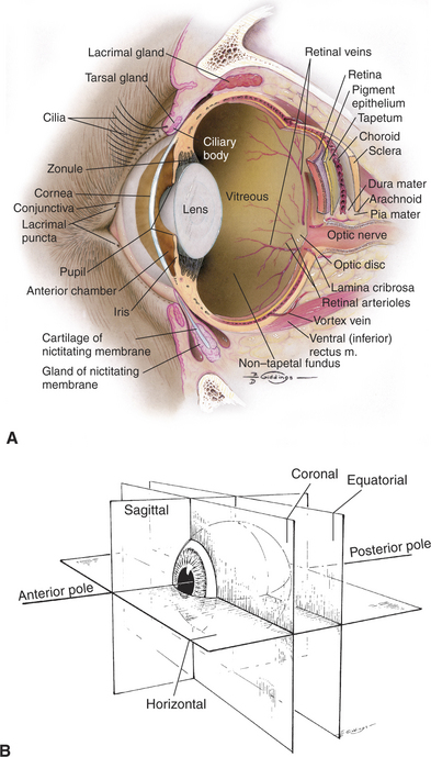
Special Senses of the Dog. The pictures in this section are reprinted with permission by the copyright owner, Hill's Pet Nutrition, from the Atlas of Veterinary Clinical Anatomy. These illustrations should not be downloaded, printed or copied except for personal, non-commercial use.

Three blind dogs
Eye Diagram Handout Author: National Eye Health Education Program of the National Eye Institute, National Institutes of Health Subject: Handout illustrating parts of the eye Keywords: parts of the eye, eye diagram, vitreous gel, iris, cornea, pupil, lens, optic nerve, macula, retina Created Date: 12/16/2011 12:39:09 PM

Help, My Dog Has Eye Boogers! – Pets Principle
Anatomy of the cat's or dog's eye. Anatomy of the cat's or dog's eye. Vertical section of the eye and eyelids. Third eyelid and Tapetum lucidum. Schematic diagram. detailed illustration. dog eye stock illustrations

Problem Solving Using the Why Tree Video
Due to hundreds of years of selective breeding and the production by man of numerous types of dog breeds, dogs have the greatest variation of eye and orbit structure of any species. For example, such breeds as the brachycephalic dogs (those with short, wide heads) have eyes that appear to be more prominent, e.g. Pekingese, Boston terrier and pug.
Cataract Surgery
The shape of the eye and its placement on the head varies with different breeds. Most are oval and placed midway between the side and front of their faces. Dog fanciers have terms to describe certain eye colors and shapes: An eye that is clear blue but flecked with a white or lighter blue is known as a China Eye.
How Does the Eye Work?
Created Date: 6/11/2019 3:30:46 PM

A: Photograph of the left eye of the dog at presentation ...
Start studying Dog eye anatomy. Learn vocabulary, terms, and more with flashcards, games, and other study tools.

Giveaway & Excerpt: How to Take Beautiful Pictures of Your Cat
Dogs therefore have two sets of teeth, baby teeth (28), which will eventually fall out, and adult teeth (42). Different types of dog teeth. As you can see in the diagram above, there are different types of dog teeth. These different types of dog teeth include Incisors, Canine, Premolars, and Molars. How many teeth do dogs have on top

Your Overtaxed Eyes and What to do About Them — Musicians ...
A bull's eye diagram is a strategic template designed in the shape of a bull's eye. It helps teams work out where their real priorities lie. Each concentric circle radiating out from the center corresponds to a different level of importance. The inner-circle should hold only the absolute top-priority tasks, all the way to the outermost ...
Phantom
Anatomy of the eye. The bony cavity or socket that contains the eyeball is called the orbit. The orbit is a structure that is formed by several bones. The orbit also contains muscles, nerves, blood vessels, and the structures that produce and drain tears. The white of the eye is called the sclera. This is the relatively tough outer layer of the ...
Blogs

Taking Care of Your Dog's Eyes | CleverPet
Eye exam chart stock image. Image of ophthalmology ...
Dog Eye Anatomy

The Many Adventures of Wonder Ruby: My Eye, My Eye...
Anatomy Rendering Gallery | Kezan's Portfolio
The Anatomy and Physiology of Animals/Special Senses ...

Frog, Deep Sea World, North Queensferry, Fife, Scotland.
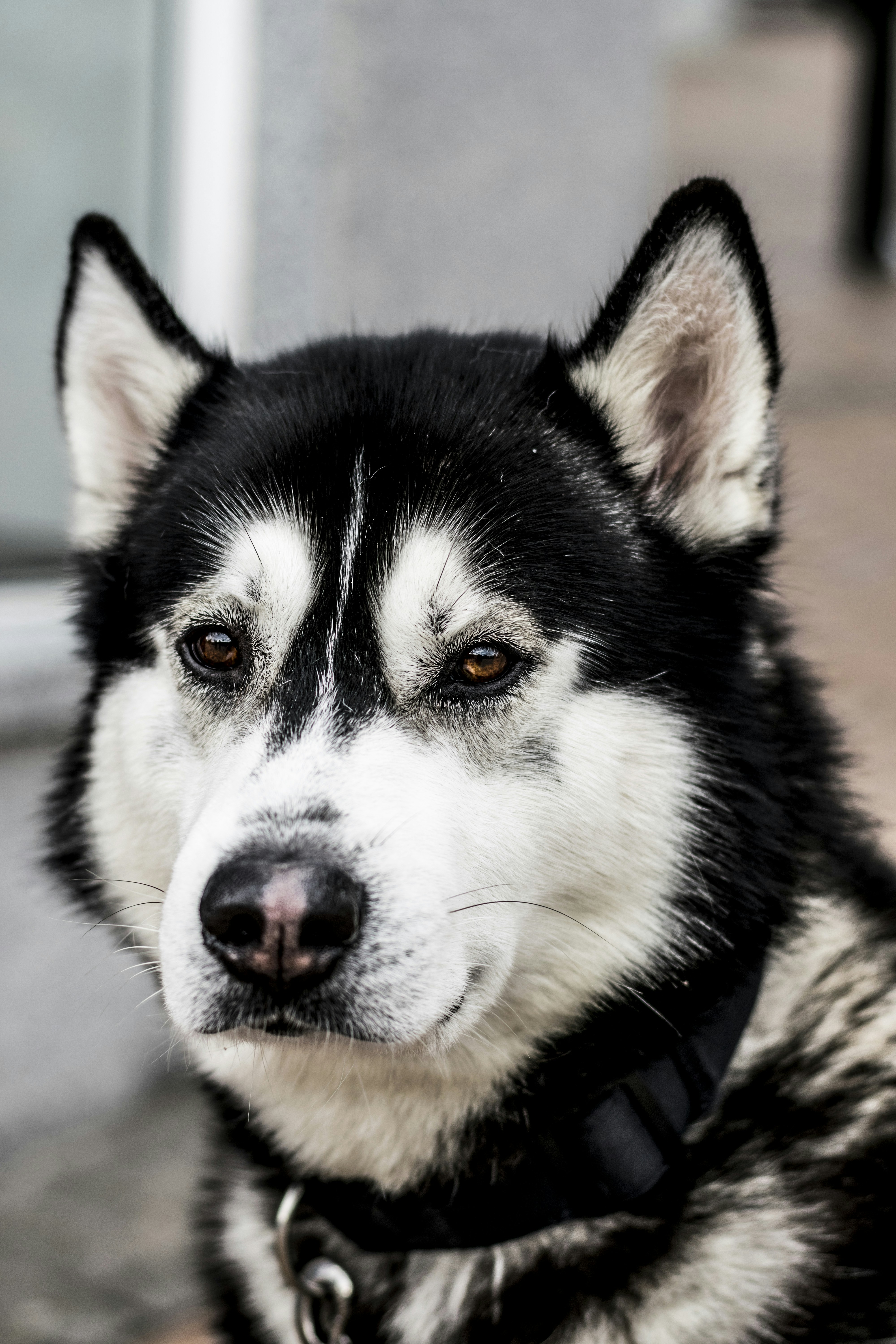
Health Issues - The Siberian Husky Club of Canada Inc

Paphos Hotel, Paphos, Republic Of Cyprus.
Structure and Function of the Eye | Veterian Key

Dog Eye Anatomy

Corneal Ulcers in Cats | VCA Animal Hospital
GC66ZT0 Sarny przy drodze (Multi-cache) in Kujawsko ...

Golden retriever dog

Eye Anatomy and Function in Animals | Dog eyes, Eye ...
Corneal Ulcers in Dogs and CatsThe Veterinary Expert| Pet ...
Blue is the New Brown – The Paw Print


Comments
Post a Comment