40 skin labeling diagram
Some sun safety tips: limit time in sun, wear clothing to cover skin exposed to the sun, use broad spectrum sunscreens with SPF values of 15 or higher regularly and as directed, reapply sunscreen ... How to: Individually Customize Axis Labels. Nov 17, 2021; 2 minutes to read; Apart from the capability to customize the overall appearance of axis labels, you can obtain individual axis labels at runtime.Then, it's possible to apply all the options available for axis labels to them, individually.
This is an online quiz called Skin Labeling. There is a printable worksheet available for download here so you can take the quiz with pen and paper.
Skin labeling diagram
a-c Label-free RCM images of three different types of ex vivo skin tissue areas, including a normal skin, b a melanocytic nevus, and c skin containing BCC, which are used as input of the virtual ... This approach may allow diagnosticians to see the overall histological features of intact skin without invasive skin biopsies or the time-consuming work of chemical processing and labeling of tissue." According to Rubinstein, this is an exciting proof-of-concept study. FPnotebook.com is a rapid access, point-of-care medical reference for primary care and emergency clinicians. Started in 1995, this collection now contains 7013 interlinked topic pages divided into a tree of 31 specialty books and 738 chapters.
Skin labeling diagram. Hair follicles are tiny holes or pores in your skin. Their main function is to grow hair. The scalp of your head too has hair follicles. In biological terms, hair follicle looks like a tunnel-shaped structure situated in the epidermis (outer layer of the skin) . Hair growth starts at the bottom of the hair follicle. Start studying Skin Structure labeling. Learn vocabulary, terms, and more with flashcards, games, and other study tools. The superficial nerves of the face and scalp are derived from three sources located in the head and neck:. Facial nerve (CN VII), which provides motor innervation to the muscles of the face; Trigeminal nerve (CN V), which provides sensory innervation to the face via its ophthalmic division (CN V1), maxillary division (CN V2) and mandibular division (CN V3) Key questions answered about coronavirus variant first detected in southern Africa First published on Fri 26 Nov 2021 07.53 EST The variant was initially referred to as B.1.1.529, but on Friday ...
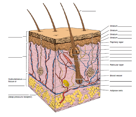
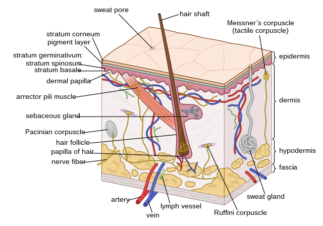
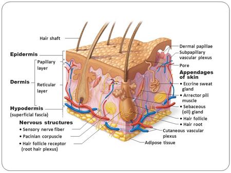
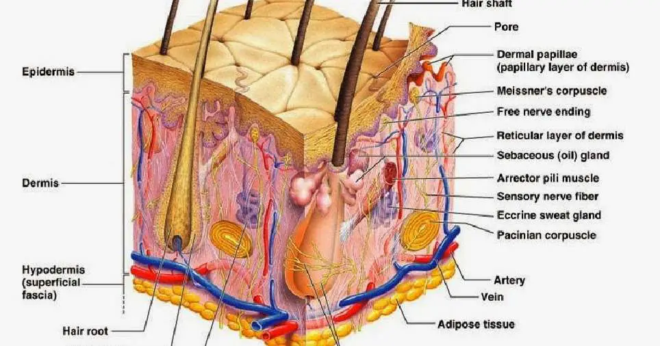
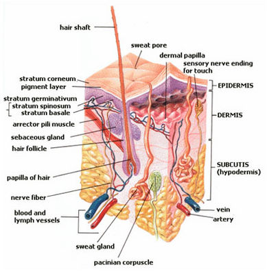
The hypodermis is the innermost (or deepest) and thickest layer of skin. It is also known as the subcutaneous layer or subcutaneous tissue . The layers of the skin include the epidermis (the outermost layer), the dermis (the next layer which is loaded with blood vessels and nerves), and then the hypodermis. 1 . (Comment 4) Several comments stated that the proposed special controls related to the disinfection of skin and/or reusable device components for single patient use blood lancets (subsets 1, 2, or 3), along with the associated special controls concerning labeling and validation, were either too burdensome or not appropriate. Nov 29, 2021 - Label The Skin Anatomy Diagram Tag Human Skin Diagram Label Human Anatomy Diagra. 21 posts related to Skin Cancer Layer Diagram. Skin Cancer Pathophysiology Diagram. Throat Cancer Diagram. Lung Cancer Labeled Diagram. Double Layer Roofing Sheet Roll Forming Machine. Pca Skin Certification Classes. ... 30603 label template; tube notch template; lease agreement;
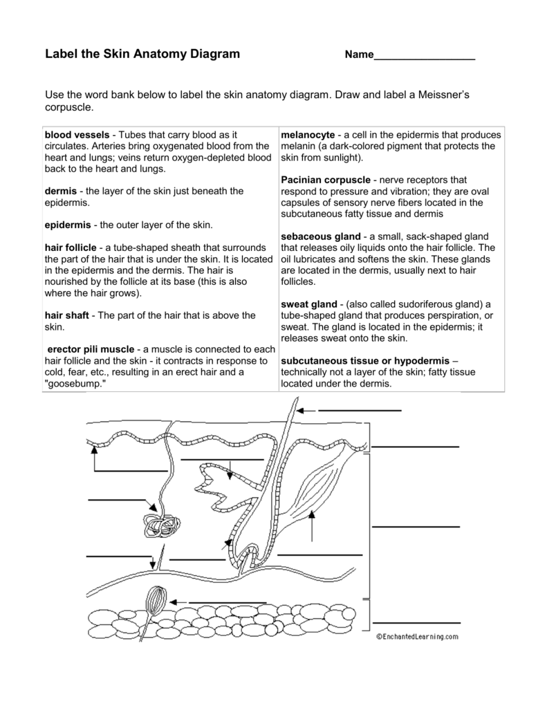
How to Teach the Muscles of the Human Body. I divide the muscle unit of my anatomy class into two sections. The first deals with the physiology of muscles, how they contract, and the tissues involved. In the second section, the focus is on the names of muscles and how they move the body. Students examine muscles of the body in sections ... The integumentary system is made up of several organs and structures including the skin, hair, nails, glands, and nerves. The primary function of the integumentary system is to protect the inside of the body from elements in the environment—like bacteria, pollution, and UV rays from the sun. The skin and its associated structures also retain ... Read the definitions, then label the skin anatomy diagram below. blood vessels - Tubes that carry blood as it circulates. Arteries bring oxygenated blood from ... Step One. Lab Four is about "Tissues" and is an introduction to Histology. We'll also look at the structure and function of the skin in the Integumentary system. Instructions: Click on the following links to view the Pre-Lab Lecture Tutorials on your introduction to tissues and the skin. Lab 4 Tutorial by Mitch Albers.
31 Label The Skin Structures And Areas Indicated In The Accompanying Labels Database 2020
Nervous system (anterior view) The nervous system is a network of neurons whose main feature is to generate, modulate and transmit information between all the different parts of the human body.This property enables many important functions of the nervous system, such as regulation of vital body functions (heartbeat, breathing, digestion), sensation and body movements.

Label The Skin Anatomy Diagram Tag Human Skin Diagram Label Human Anatomy Diagra In 2021 Skin Anatomy Integumentary System Anatomy Organs
Skin Search 4) Ishikawa Diagram for Presentations Ishikawa diagram is a helpful and effective tool when presenting specific issues and solutions to your team. With it, you can use it to present a particular outcome and the better ways to attain it, discover solutions, or deconstruct an issue.

Associate Degree Nursing Physiology Review Skin Anatomy Skin Structure Plexus Products
Suppliers and employers must use and follow the WHMIS 2015 requirements for labels and safety data sheets (SDSs) for hazardous products sold, distributed, or imported into Canada. Please refer to the following OSH Answers documents for information about WHMIS 2015: WHMIS 2015 - General. WHMIS 2015 - Labels.
Skin Diagram To Label Labelled Diagram
The ulnar nerve is a terminal branch of the medial cord of the brachial plexus.It contains mainly fibers from the anterior rami of spinal nerves C8 and T1, but may sometimes carry C7 fibers as well. From its origin, the ulnar nerve courses distally through the axilla, arm and forearm into . It is a mixed nerve and provides motor innervation to various muscles of the forearm and ...

Given Below Is A Diagrammatic Sketch Of The Vertical Section Of The Human Skin A Label The Parts Numbered From 1 To 9 B State One Main Function Of Each Of The Following Parts Part
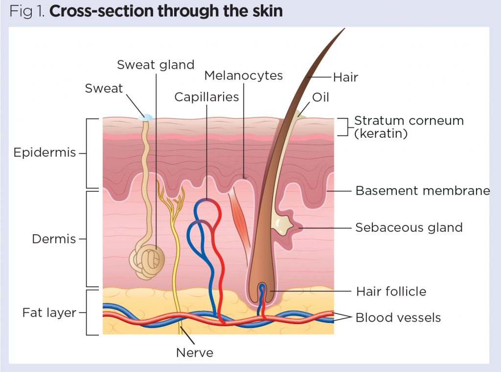
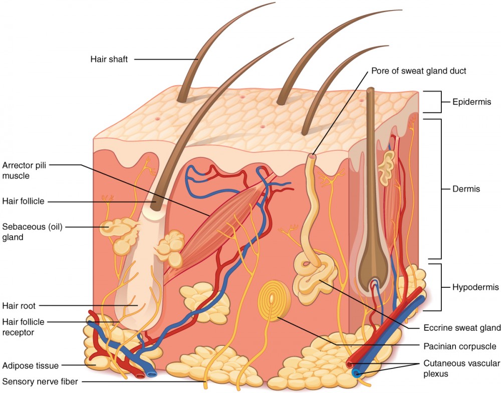
The skin is made up of 3 layers. Each layer has certain functions: Epidermis. Dermis. Subcutaneous fat layer (hypodermis) ...
Human Skin Wikipedia
This approach may allow diagnosticians to see the overall histological features of intact skin without invasive skin biopsies or the time-consuming work of chemical processing and labeling of tissue."
Skin Diagram Let S Label It Up Skin Diagram Left Epidermis Dermis Hypodermis Subcutaneous Ppt Download
No information is available for this page.Learn why

The Skin The Eye And The Ear Musculoskeletal Key
The melanin is transferred into the keratinocytes via a cellular vesicle called a melanosome (Figure 7). This figure consists of two diagrams side by side. The ...

Skin Anatomy Cross Section With Labels On White Stock Photo Download Image Now Istock
The dermis is the middle layer of the three layers of skin. It's located between the epidermis and the subcutaneous tissue. It contains connective tissue, blood capillaries, oil and sweat glands, nerve endings, and hair follicles. The dermis is split into two parts—the papillary dermis, which is the thin, upper layer, and the reticular dermis ...

33 Label The Structure Of The Skin Label Design Ideas 2020
Brainstem tectum, tegmentum and basal area (diagram) The tectum is the roof of the cavity while the tegmentum forms the ventral covering.The central cavity of the neural tube becomes the aqueduct of Sylvius, the fourth ventricle, and the central canal of the spinal cord.Therefore the tectum is the area dorsal to the aqueduct of Sylvius (in the midbrain) and fourth ventricle (at the pons ...

Sectioned Sebaceous Human Anatomy Guws Medical
Cumberland Pharmaceuticals Inc. (NASDAQ: CPIX), a specialty pharmaceutical company today announced the U.S. Food and Drug Administration (FDA) has approved expanded labeling for Caldolor®, an ...
Skin 1 The Structure And Functions Of The Skin Nursing Times
After learning, comparing and contrasting the steps of the engineering design process (EDP) and scientific method, students review the human skeletal system, including the major bones, bone types, bone functions and bone tissues, as well as other details about bone composition. Students then pair-read an article about bones and bone growth and compile their notes to summarize the article.

Pin On Adobe Stock Photos For Sale
Type of ingredient: Humectant. Main benefits: Improves dryness, replenishes the skin, and provides structure and volume. Who should use it: It is recommended for people of all ages and skin types and is especially beneficial for those with dry, dehydrated skin. How often can you use it: Sodium hyaluronate is safe to use in concentrations of up to 2% twice daily, morning and night.

Image Of The Human Skin Learn More About Your Skin S Structure And Functions Subcutaneous Tissue Skin Structure Epidermis
Capillary malformation (arteriovenous malformation syndrome) may occur as part of an inherited syndrome present in roughly 1 in 100,000 people of European ancestry. In this syndrome, there is more blood flow than normal through the capillaries near the skin, which results in pink and red dots on the skin.

Skin Structure Labeling Diagram Quizlet
Skin Diagram Labeling. 1. Label the diagram with the letters below according to the structure/area they describe. You may label with a line or put the label ...3 pages

Label The Skin Quiz
Skin diagram unlabeled. You know what's coming next. It's time to label the diagram for yourself! Click below to download a free unlabeled version of the ...
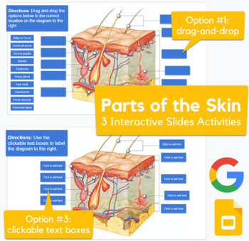
Skin Diagram Drag And Drop Labeling Activity In Slides Advanced
The skin allows for bodily growth and adapts to suit an individuals course of movement. It has elastic and recoil properties on all of its layers, meaning it can adapt for growth and movement. 6. Excretion. The skin can also expel uric acid, ammonia, urea, and excess water. When noting the functions of the skin, excretion is a very important one.

32 Label These Structures Of The Integumentary System Labels Database 2020
The brachial plexus is a network of nerves that gives rise to all the motor and sensory nerves of the upper extremity his plexus arises from the anterior rami of spinal nerves C5-T1 that undergo several mergers and splits into trunks and divisions, until they finally give rise to their terminal branchesThese terminal branches are responsible for motor and sensory innervation of the upper ...
Label The Skin Anatomy Diagram
Symptoms of imbalance: Menstrual disorders, dry skin and eyes, jaundice, blurred vision, vertigo, stiff joints, and headaches. On an emotional level, an imbalance of energy in the liver meridian results in anger, irritability, depression, and a lack of control and emotional flexibility. Time of day: 1:00am-3:00am.

Anatomy Of Human Skin Cross Section View Labeled On White Stock Photo Download Image Now Istock
Basic structure of the skin. A basic understanding of skin anatomy is important when explaining the process of. The Skin Blank Diagram Pdf from imgv2-2-f.scribdassets.com Dead as it may be, your skin cells still need proper upkeep. This poster clearly labels the:. Browse skin layers resources on teachers pay teachers,.

Label The Skin Quiz
FPnotebook.com is a rapid access, point-of-care medical reference for primary care and emergency clinicians. Started in 1995, this collection now contains 7013 interlinked topic pages divided into a tree of 31 specialty books and 738 chapters.
.jpg)
A Human Body Skin Structure Quiz Proprofs Quiz
This approach may allow diagnosticians to see the overall histological features of intact skin without invasive skin biopsies or the time-consuming work of chemical processing and labeling of tissue." According to Rubinstein, this is an exciting proof-of-concept study.

Functions Of The Integumentary System Boundless Anatomy And Physiology
a-c Label-free RCM images of three different types of ex vivo skin tissue areas, including a normal skin, b a melanocytic nevus, and c skin containing BCC, which are used as input of the virtual ...
Skin Diagram Labeled

Skin Diagram Worksheet Education Com

Chapter 4 Xlsx Exercise 4 2 Labeling The Skin Using The Following List Choose The Correct Terms To Label The Diagram Correctly Adipose Tissue Artery Course Hero

Imagequiz Labeling Skin Diagram 2016

89 Skin Diagram To Label Illustrations Clip Art Istock

Quia Class Page 5th Period
Layers Of The Skin Anatomy And Physiology I
Integumentary System Histology Illustrations Skin Labels Histology Illustrations

34 Skin With Label Labels Design Ideas 2020

A Diagrammatic Representation Of The Structure Of Human Skin In Cross Download Scientific Diagram
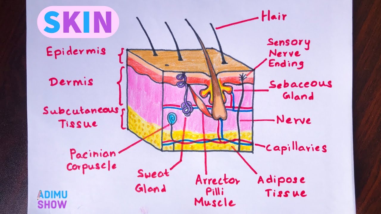
How To Draw Skin Layers Integumentary System Step By Step Drawing Youtube

Skin Diagram And Information About Your Skin Skin Anatomy Integumentary System Psoriasis Skin

Skin Labeling Quiz

Skin Diagram With Detailed Illustrations And Clear Labels

Cross Section Human Skin Labels Stock Illustration 14728675
3

Layers Of Skin How Many Diagram Model Anatomy In Order


Comments
Post a Comment