39 brain anatomy diagram sagittal
Illustrations: Sagittal view of brain with lobes and general anatomy labeled. Diagrams. Thumbnails: File under medical illustrations showing Sagittal View of Brain, with emphasis on the terms related to anatomy sagittal brain lobe corpus callosum thalamus midbrain pons medulla oblongata cerebellum. The coronal plane, horizontal plane and sagittal plane are shown in the figure ... The figures below show the human brain in the three planes of section on ...
Download scientific diagram | Brain regions anatomy. Sagittal section of adult mice brain. From panel a to c sagittal sectioning from the sagittal superior sinus toward the border of the cerebral ...

Brain anatomy diagram sagittal
• Sectional Anatomy of the Brain • Sectional Anatomy of the Spine Outline Slide # 3 Upon completion of this course, the attendee should… 1. Learn about planes of the brain & spine 2. Learn sectional anatomy of the brain Para Sagittal 3. Learn sectional anatomy of the spine Part I Objectives Slide # 4 Axial PDWI Midline Sagittal T1WI ... Figure 1.14Midsagittal view of the human brain. ... The other prominent anatomical feature of the midbrain—the cerebral peduncles (also visible from the ... Brain Diagram Sagittal View. angelo. November 26, 2021. Vertical Section Of A Human Brain Showing The Medulla Pons Cerebellum Hypothalamus Thalamus Midbrain Stock Vector Human Brain Human Brain Diagram Brain. Mid Sagittal Section Through The Human Brain By Destroma Deviantart Com On Deviantart Human Brain Diagram Brain Diagram Human Brain.
Brain anatomy diagram sagittal. 20/07/2020 · The paired left and right transverse sinuses are major dural venous sinuses and arise from the confluence of the superior sagittal, occipital and straight sinuses at the torcular herophili (confluence of sinuses).. On each side, the transverse sinus then runs in the lateral border of the tentorium cerebelli and grooves the occipital and squamous temporal bones. 26/09/2021 · The trigeminal nerve, also known as the fifth (or V) cranial nerve, is a cranial nerve and its primary role is relaying sensory information from the face and head, although it does provide motor control to the muscles of mastication.It is both large and complicated and has multiple brainstem nuclei (sensory and motor) as well as many interconnections with other … Jan 19, 2020 — Media in category "Human brain (sagittal section)". The following 156 files are in this category, out of 156 total. Start studying Structures of the Brain - Sagittal Section. Learn vocabulary, terms, and more with flashcards, games, and other study tools.
Find labeled brain anatomy stock images in HD and millions of other royalty-free stock ... Human Brain Anatomy Sagittal Section with Labels, 3D Rendering. The Brain - Sagittal Section. Create healthcare diagrams like this example called The Brain - Sagittal Section in minutes with SmartDraw. SmartDraw includes 1000s of professional healthcare and anatomy chart templates that you can modify and make your own. 5 Sectional Planes Used in Anatomy • Coronal: taken in the plane of the face • Sagittal: taken in the plane dividing the 2 hemispheres, perpendicular to the coronal plane • Horizontal (axial transverse): taken parallel to the rostral-caudal axis of the brain; so if an individual is standing upright, these sections are Sagittal section of the brain. Human brain internal anatomy vector diagram. Sagittal section of the brain. Medical infographic
Dural Venous Sinuses Of The Brain Diagram. In this image, you will find superior sagittal sinus, falx cerebri, inferior sagittal sinus, straight sinus, cavernous sinus, transverse sinuses, sigmoid sinus, jugular foramen, right internal jugular vein in it. Health care advices from Overseas Doctor . We are pleased to provide you with the picture ... Simply a cut of the human brain in a sagittal view and an extra image in case you want to learn some anatomy. -RSB [via here & here) Related Stories:Felted AnatomyPaper AnatomySaturn V Cutaway by Stephen BiestyDan QuintanaWire AnatomyColored Anatomy Plates from Essai D'Anatomie - 1745Golf Ball Cross-Sections by James FriedmanA Soft and Squishy BrainZemanta Your Thoughts Are Welcome Spinal cord. it connects a large part of the PNS to the brain. Gyrus. increases the amount of cerebral cortex that can fit in the skull. Brainstem. controls the flow of messages between the brain and the rest of the body, and it also controls basic body functions such as breathing, swallowing, heart rate, blood pressure, consciousness, and ... Figure 7-4 is a diagram of the sagittal view of the human brain. First, match the letters on the diagram with the list of terms and insert the appropriate letter in each answer blank. Then, color the brain-stem areas blue and the areas where cerebrospinal fluid is found yellow. 1. Cerebrum 8. Pituitary gland (Endocrine gland called Master Gland) 2.
File Human Brain Sagittal Section Svg Wikimedia Commons
Vertical Section Of A Human Brain Showing The Medulla Pons Cerebellum Hypothalamus Thalamus Midbrain Stock Vector Human Brain Human Brain Diagram Brain. Sheep Brain Sagittal Section Medial View Brain Anatomy Sheep Brain In 2021 Brain Anatomy Sheep Nervous System Anatomy. Image Not Available Brain Anatomy Brain Anatomy And Function Human Brain ...

1 Schematic Overview Of Basic Brain Anatomy In A Sagittal Plane Download Scientific Diagram
CROSS SECTIONAL ANATOMY OF BRAINSOURCE: MRIMASTER.COM
Human Brain Anatomy Sagittal Plane Pituitary Gland Png Clipart Abdomen Brain Brainstem Flesh Frontal Lobe Free
Our interactive diagram helps you explore the anatomy of the human brain and learn all about how it functions. Skip Navigation. ... Anatomy of the Brain. ... The superior sagittal sinus is a vein that runs through the longitudinal fissure of the brain and carries blood and cerebrospinal fluid from the brain back to the heart.

Sagittal Cut Brain Anatomy Diagram Quizlet
Anatomy of the Brain Overview. The brain is an amazing three-pound organ that controls all functions of the body, interprets information from the outside world, and embodies the essence of the mind and soul. Intelligence, creativity, emotion, and memory are a few of the many things governed by the brain.
Color Coded Human Sagittal Cut Half Skull With Brain Hemisphere Bone Clones Inc Osteological Reproductions
Three anatomical sections of the brain (axial, coronal and sagittal) close this chapter on the brain. Brain , Coronal section : Brain , Anatomy diagram. Numerous illustrations are available on the cerebellum, representation of cerebellar lobes, fissures, sulci and the vermis. Cerebellum , Anterior view : Diagram.

Human Brain Anatomy Composition 3468357 Vector Art At Vecteezy
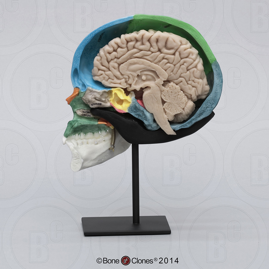
The ethmoid bone (/ ˈ ɛ θ m ɔɪ d /; from Greek ethmos, "sieve") is an unpaired bone in the skull that separates the nasal cavity from the brain.It is located at the roof of the nose, between the two orbits.The cubical bone is lightweight due to a spongy construction. The ethmoid bone is one of the bones that make up the orbit of the eye.
Brain Resource Imageshare
The cerebellar view is angled through the posterior fossa (cisterna magna). All are axial (transverse) views of the fetal head with the exception of the cerebellar view, which is axial oblique. Other planes include the sagittal, parasagittal, and coronal which may be useful or necessary to define anatomy. Thalamic View. Above.
Duke Neurosciences Lab 1 Surface Anatomy Of The Brain
Brain Model, sagittal view « Inferior view | Brain main | Inferolateral view ». Use the diagram below to: Identify the labeld structures (A through S & i through ix)

Sagittal Brain Anatomy Diagram Quizlet
We are pleased to provide you with the picture named Cerebrum Brain In Situ Sagittal Section Medial Section.We hope this picture Cerebrum Brain In Situ Sagittal Section Medial Section can help you study and research. for more anatomy content please follow us and visit our website: www.anatomynote.com. Anatomynote.com found Cerebrum Brain In Situ Sagittal Section Medial Section from plenty of ...

Sagittal Section Of Human Brain Diagram Quizlet
09/06/2020 · Sagittal Plane. Cuts or divides the body into right and left portions. ... This diagram will be very useful as it provides a visual guide on how the blood flows via the heart and its major structures. ... The nervous system consists of the anatomy of the brain and the communication between neurons and other parts of the human body.

Cross Sectional Anatomy Mri Brain Sagittal Anatomy Free Mri Brain Cross Sectional Anatomy
The module on the anatomy of the brain based on MRI with axial slices was redesigned, having received multiple requests from users for coronal and sagittal slices. The elaboration of this new module, its labeling of more than 524 structures on 379 MRI images in three different views and on 26 anatomical diagrams, took more than 6 months.
481 Thalamus Vector Images Thalamus Illustrations Depositphotos
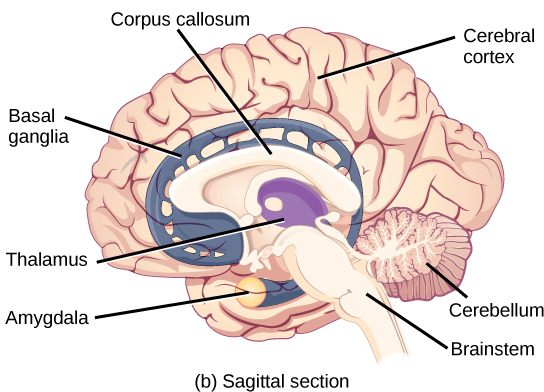
BI 335 – Advanced Human Anatomy and Physiology Western Oregon University Figure 4: Mid-sagittal section of brain showing diencephalon (includes corpus callosum, fornix, and anterior commissure) Marieb & Hoehn (Human Anatomy and Physiology, 9th ed.) – Figure 12.10 Exercise 2: Utilize the model of the human brain to locate the following structures / landmarks for the
2
This MRI brain cross sectional anatomy tool is absolutely free to use. Use the mouse scroll wheel to move the images up and down alternatively use the tiny arrows (>>) on both side of the image to move the images.>>) on both side of the image to move the images.

Sagittal Section Of Human Brain With Labeled Parts Stock Photo Download Image Now Istock
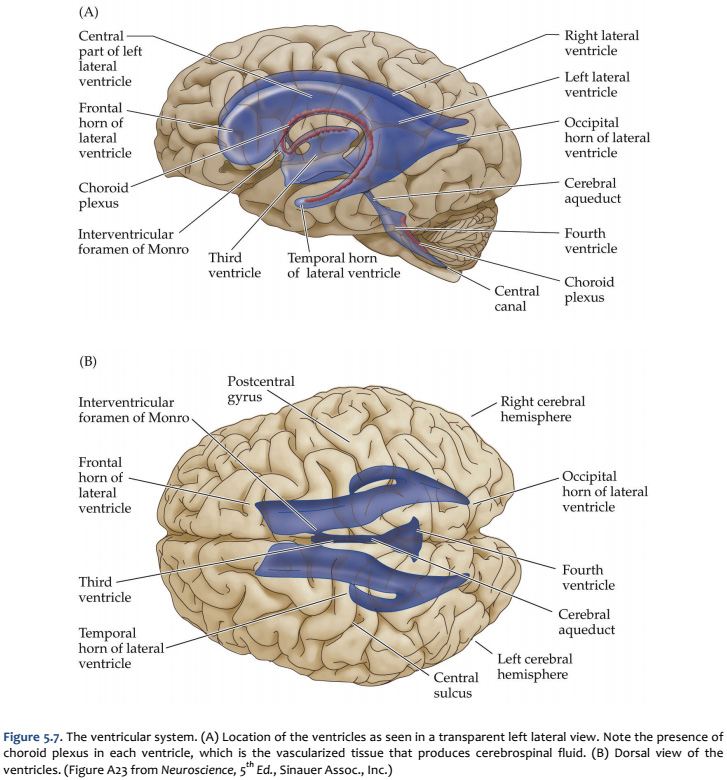
Learn the ventricles of the brain along with their definition, function, location, anatomy, and cerebrospinal fluid (CSF) flow using labeled diagrams. The ventricular system contains the lateral, third, and fourth ventricles whose function is to produce cerebrospinal fluid. Learn where CSF is found,
Anatomical Diagram Brain Images Stock Photos Vectors Shutterstock
Veins of the Sagittal Brain. Create healthcare diagrams like this example called Veins of the Sagittal Brain in minutes with SmartDraw. SmartDraw includes 1000s of professional healthcare and anatomy chart templates that you can modify and make your own.

Human Brain Detailed Anatomy Stock Vector Illustration Of Cerebellum Chart 54989259
Neuroanatomy Tutorial - Labeled Images. This tutorial has images in which the structures are labeled. You are to identify the structures by clicking on the name of the structure. The structure whose name is clicked will be identified in the image by an arrow.

Figure The Fore Brain Or Prosencephalon Mesal Statpearls Ncbi Bookshelf
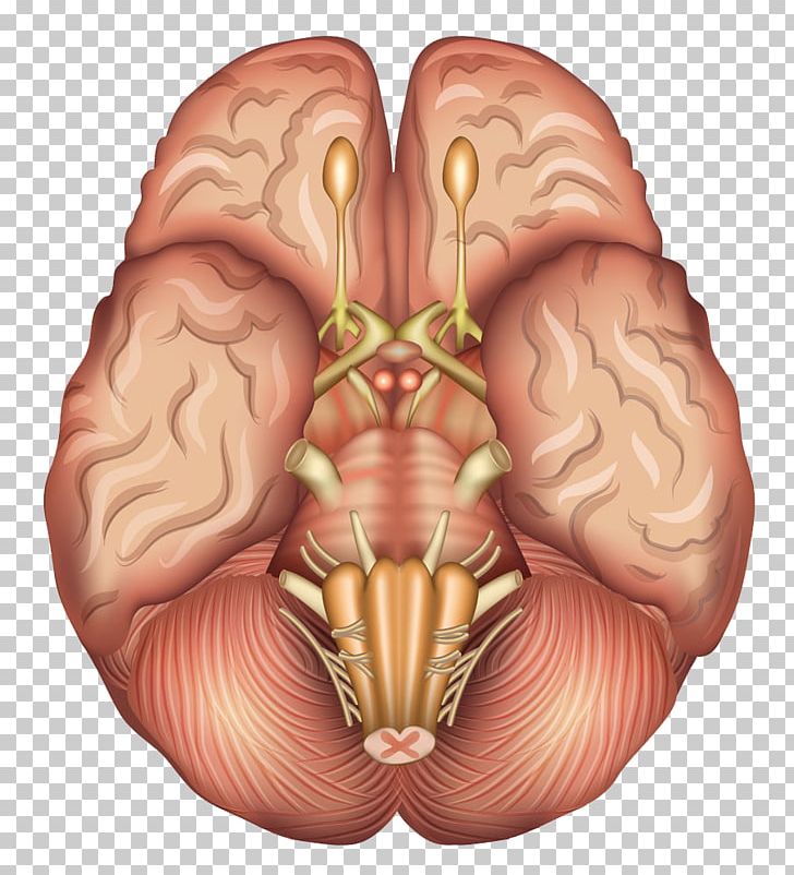
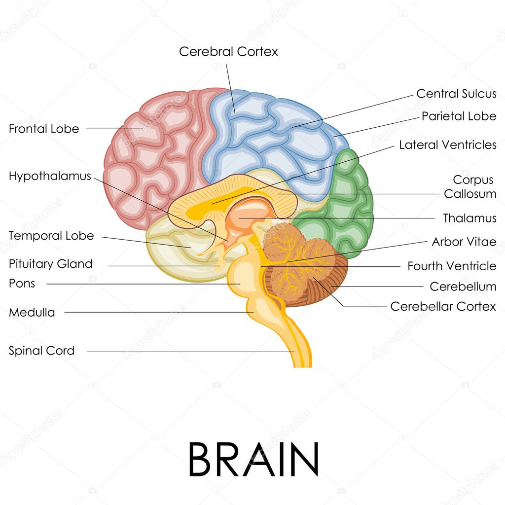
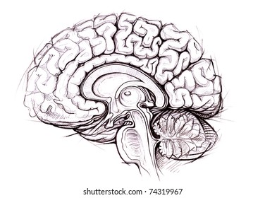
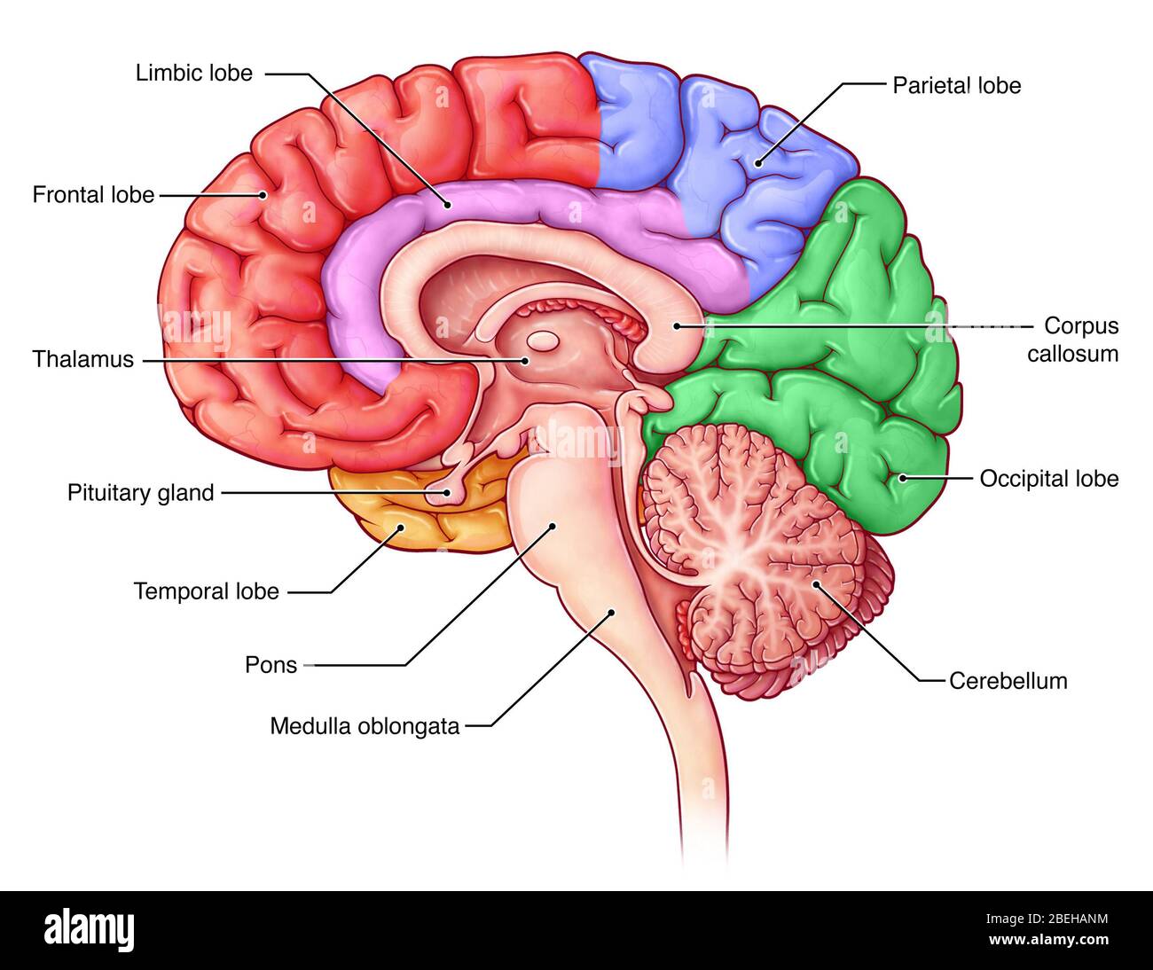
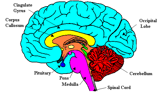
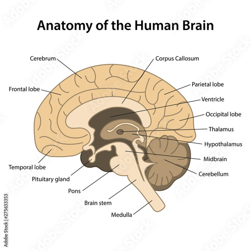
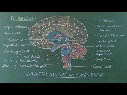
The midsagittal section of the brain shows the three major parts of the brain, which are the cerebrum, cerebellum, and brainstem.These brain parts are marked with visible gross features like the gyri (singular: gyrus) and sulci (singular: sulcus) of the cerebrum. They are each also divided into subparts or regions for simplified localization of structures, for example, the brainstem is ...
Midsagittal Section Of The Brain Anatomy Kenhub
Lobes of the brain: A diagram of the brain identifying the different lobes by color. Counterclockwise from bottom: It contains the parietal lobe (green), the occipital lobe (red), the temporal lobe (yellow), and the frontal lobe (blue).
Lobes Of The Brain Sagittal View Stock Photo Alamy
Illustration about Human brain internal anatomy vector diagram. Sagittal section of the brain. Medical infographic. Illustration of sagittal, medical, anatomy - 204571373
Neuroscience For Kids The Brain Right Down The Middle
Welcome to Soton Brain Hub- videos to help explain the mysteries of the brain!In her first video for Soton Brain Hub Ellie describes the anatomy of the brain...

Human Brain Anatomy Set Of Lateral Sagittal Superior Inferior Views With All Lobes Royalty Free Cliparts Vectors And Stock Illustration Image 71810394
Brain Diagram Sagittal View. angelo. November 26, 2021. Vertical Section Of A Human Brain Showing The Medulla Pons Cerebellum Hypothalamus Thalamus Midbrain Stock Vector Human Brain Human Brain Diagram Brain. Mid Sagittal Section Through The Human Brain By Destroma Deviantart Com On Deviantart Human Brain Diagram Brain Diagram Human Brain.

Brain Sagittal Section Gray S Anatomy Illustration Radiology Case Radiopaedia Org
Figure 1.14Midsagittal view of the human brain. ... The other prominent anatomical feature of the midbrain—the cerebral peduncles (also visible from the ...

Mid Sagittal Cross Section Of Brain Brain Anatomy Brain Anatomy And Function Anatomy
• Sectional Anatomy of the Brain • Sectional Anatomy of the Spine Outline Slide # 3 Upon completion of this course, the attendee should… 1. Learn about planes of the brain & spine 2. Learn sectional anatomy of the brain Para Sagittal 3. Learn sectional anatomy of the spine Part I Objectives Slide # 4 Axial PDWI Midline Sagittal T1WI ...

108 Sagittal Vector Images Sagittal Illustrations Depositphotos

Sagittal Section Of The Brain Stock Vector Illustration Of Sagittal Medical 204571373
Brain Cross Section Anatomy Anatomy Drawing Diagram
Anatomy Of The Human Brain Sagittal Cut Structure Of The Human Brain With Main Parts Labeled Vector Illustration Stock Vector Adobe Stock
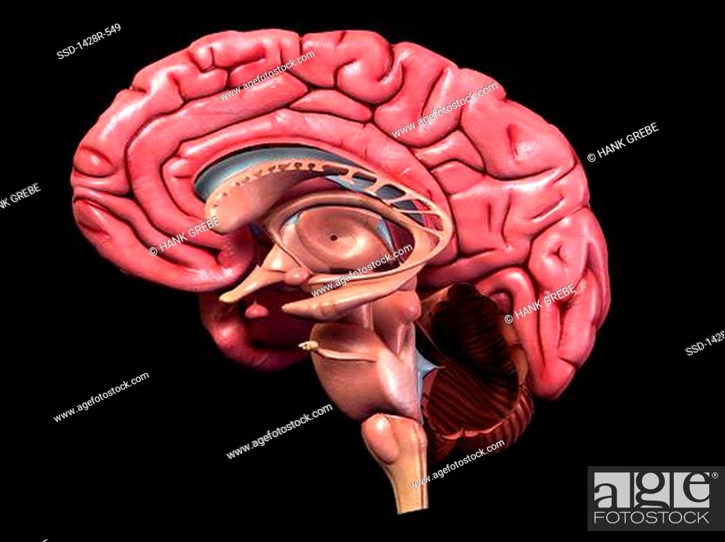
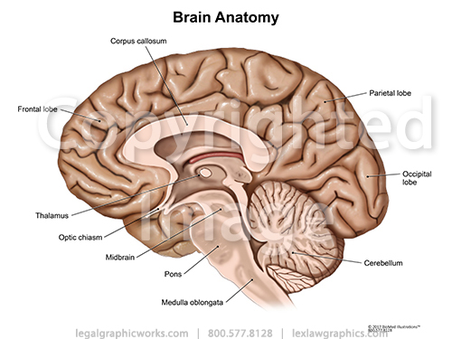
Full Color Detailed Human Brain Anatomy 3 D Medical Illustration Of Sagittal Section Side View Stock Photo Picture And Royalty Free Image Pic Ssd 1428r 549 Agefotostock

Brain Regions Anatomy Sagittal Section Of Adult Mice Brain From Panel Download Scientific Diagram
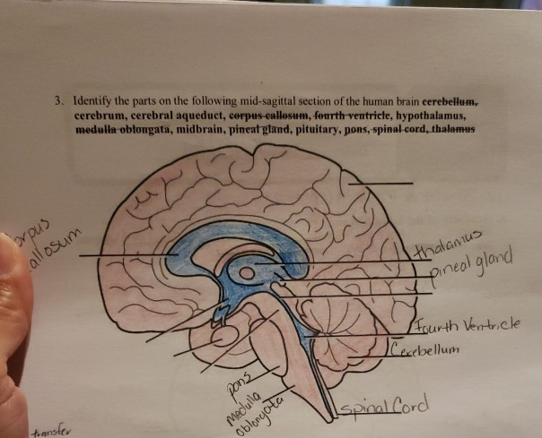
Solved 3 Identify The Parts On The Following Mid Sagittal Chegg Com
How To Draw Sagittal Section Of Human Brain Easy Steps Tricks To Draw Yo Yoo Bio Yo Yoo Diagrams Youtube

Sagittal Brain Anatomy Diagram Quizlet
3
Biorender Life Science Icons

Sagittal Section Of The Human Brain Structure Of The Human Brain Human Anatomy Medical 3d Vector Illustration Isolated On White Background Stock Illustration Download Image Now Istock

Mid Sagittal Section Through The Human Brain By Destroma Deviantart Com On Deviantart Human Brain Diagram Brain Diagram Human Brain

The Brain Stem And The Cerebelleum Human Anatomy And Physiology Lab Bsb 141

Anatomy Of Human Head Sagittal Section Download Scientific Diagram
1


Comments
Post a Comment