38 foot nerve diagram
A neuroma is a thickening of nerve tissue that may develop in various parts of the body. The most common neuroma in the foot is a Morton's neuroma, which occurs between the third and fourth toes. It is sometimes referred to as an intermetatarsal neuroma. Intermetatarsal describes its location in the ball of the foot between the metatarsal bones. The sciatic nerve is the largest and longest nerve in the human body, originating at the base of the spine and running along the back of each leg into the foot. 1, 2 At its thickest point, it is about as wide as an adult thumb. The sciatic nerve is formed in the lower spine by the combination of motor and sensory fibers from spinal nerves L4 to S3.
19/01/2021 · The tibial nerve is a major peripheral nerve of the lower limb. It has several cutaneous and motor functions in the leg and foot. In this article, we shall look at the anatomy of the tibial nerve – its anatomical course, functions and clinical correlations.
Foot nerve diagram
normal anatomy and common variants of the nerves of the foot and ankle, with use of dissected specimens and correlative US and MR imaging findings. In addition, the authors illustrate proper probe positioning, which is essential for visualizing the nerves at US. The authors' discussion focuses on the superficial and deep Nerve pain in foot can stem from two places. The Spine: Damage to the lower back is a common cause of nerve pain in the foot. This tends to cause back, buttock and leg nerve pain as well as foot pain and weakness. The Nerve: a peripheral foot neuropathy is caused by damage along the course of the nerve somewhere down the leg or in the foot itself. Problems with nerves in the feet are very common. Many times, an injured nerve will cause intense pain and heat felt within the foot. Nerves act as a network, communicating important information from the foot to the brain. Learn more about the various conditions and problems that can affect the nerves in the foot.
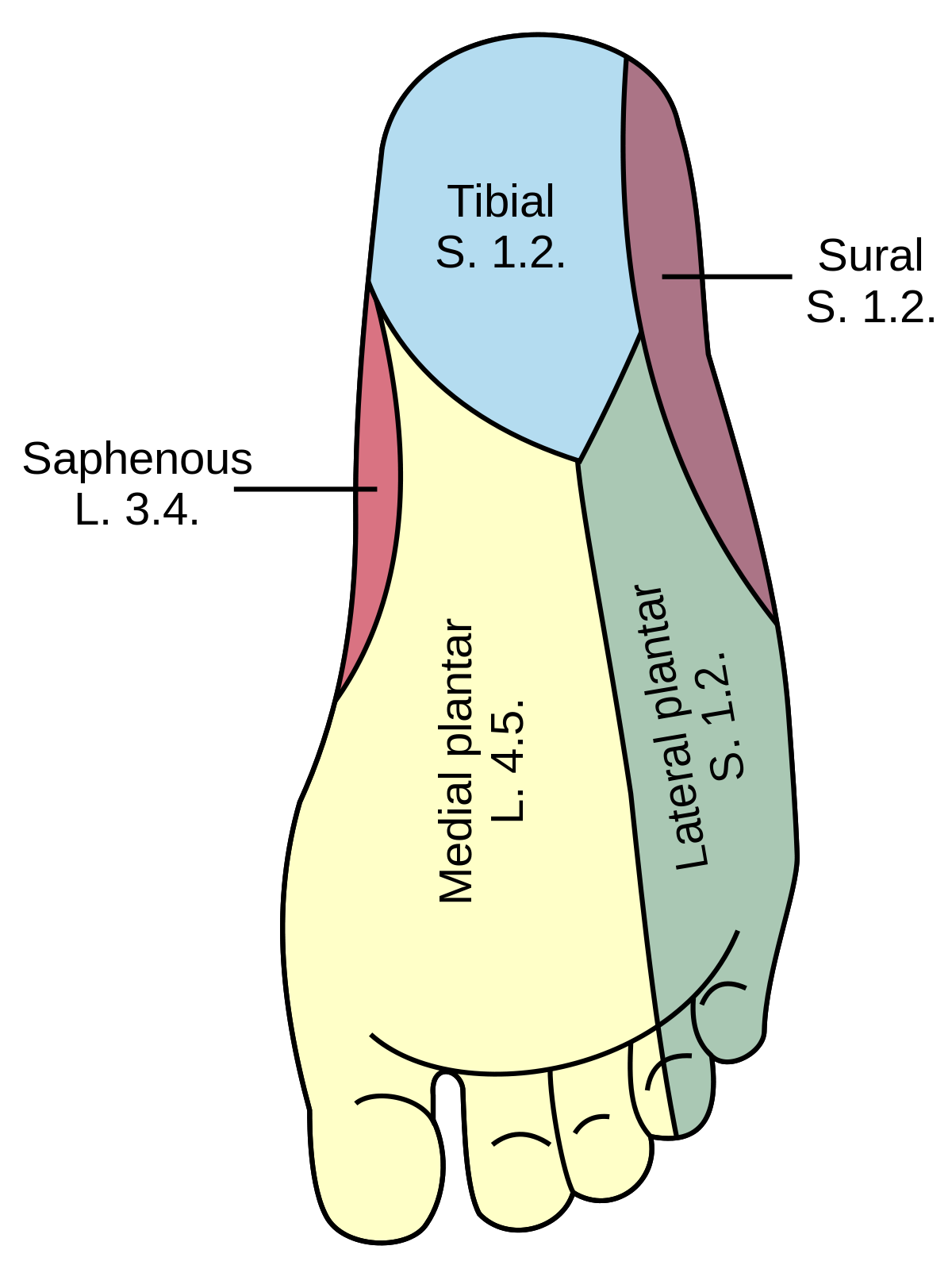
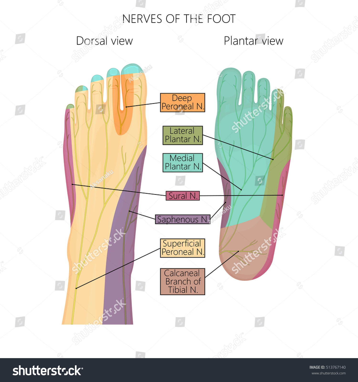
Foot nerve diagram. Foot and ankle surgeons treat all conditions affecting the foot and ankle, from the simple to the complex, in patients of all ages including diabetes. Their intensive education and training qualify foot and ankle surgeons to perform a wide range of surgeries, including any surgery that may be indicated for diabetic foot care. The lateral plantar nerve is an important motor nerve in the foot because it innervates: All intrinsic muscles in the sole, except for the muscles supplied by the medial plantar nerve. It also innervates a strip of skin on the lateral side of the anterior two-thirds of the sole. The adjacent plantar surfaces of the lateral one and one-half digits. Saphenous Nerve It is the largest cutaneous branch of the femoral nerve. It supplies cutaneous branches to the skin of the leg and foot in the region between the knee and the ankle. Sciatic Nerve Also known as the ischiatic nerve, the sciatic nerve is a nerve fiber that begins in the lower back and ends in the lower limb. 10/03/2021 · Nerve damage frequently causes burning, pins and needles, numbness and weakness in the feet. The nerve may be damaged anywhere between the lower back and the toes. Symptoms may be felt in both feet. Full Article: Nerve Pain In Foot
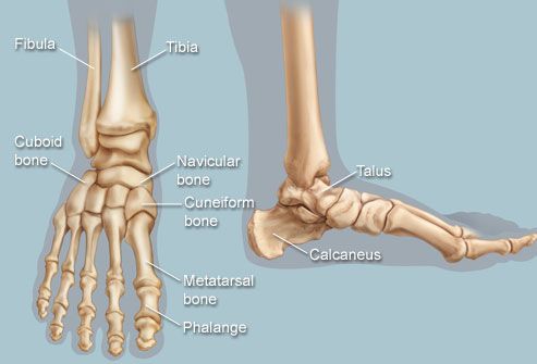
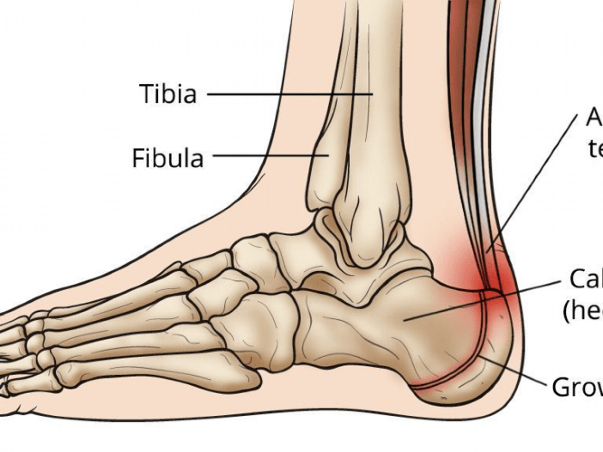
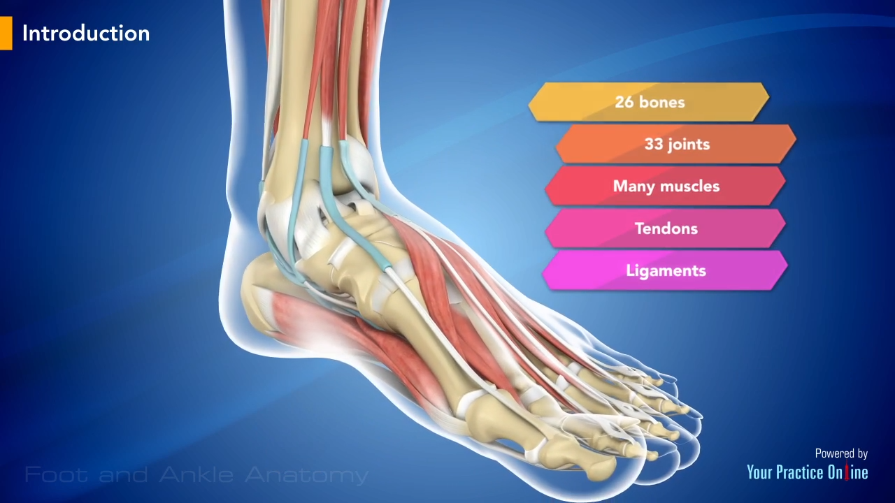
It is NOT meant to be used as a diagnostic tool, and it is NOT a substitute for a doctor's or foot specialist's care. Be sure to consult with your primary care physician or foot specialist if you have pain, discomfort, or any other symptoms of foot issues or conditions. If you feel no pain/sensation, you may have neuropathy and should see ... Spinal nerves (diagram) Spinal nervesemerge from the segments of the spinal cord. They are numbered according to their specific segment of origin. Hence, the 31 pairs of spinal nerves are divided into 8 cervical pairs, 12 thoracic pairs, 5 lumbar pairs, 5 sacral pairs, and 1 coccygeal spinal nerve. All spinal nerves are mixed, containing both ... On the chart below you will see 4 Columns (Vertebral Level, Nerve Root, Innervation, and Possible Symptoms). Under 'Vertebral Level': C1-C7 is the NECK, ; T1-T12 is the UPPER BACK/rib cage area, and ; L1-L5 is the LOWER BACK.; Simply line up the "Vertebral Level" with the "Possible Symptoms" and you will see some surprising connections of symptoms that relate to your spine. The anatomy of the foot. The foot contains a lot of moving parts - 26 bones, 33 joints and over 100 ligaments. The foot is divided into three sections - the forefoot, the midfoot and the hindfoot. The forefoot. This consists of five long metatarsal bones and five shorter bones that form the toes (phalanges). The midfoot.
by M De Maeseneer · 2015 · Cited by 70 — The normal anatomy and common variants of nerves of the foot and ankle are discussed with use of dissected specimens and correlative ... Browse 27 sciatic nerve diagram stock photos and images available, or start a new search to explore more stock photos and images. male nervous system, illustration - sciatic nerve diagram stock illustrations. engraved antique, anatomy of the brain and nerves engraving antique illustration, published 1851 - sciatic nerve diagram stock illustrations. Match the corresponding numbers on the foot diagram below for a list of conditions that may be causing your foot and ankle pain. This is meant for educational purposes only. If you're having a problem with your foot or ankle, visit a podiatrist - a foot and ankle specialist! Top (Dorsal) View of Foot & Ankle Number 1 and 2: Nerves In Foot Diagram. nerves of the leg and foot along its route through the legs the sciatic nerve splits into the tibial and mon fibular peroneal nerves which in turn split into many smaller nerves in the legs and feet the nerves of the foot help move the body and keep balance both while it's moving and at rest a plete guide to the nerves in your feet foot vitals tingling feet the ...
Feet Human Anatomy Bones Tendons Ligaments And More
A high arch is the opposite of a flat foot, and somewhat less common. The term pes cavus encompasses a broad spectrum of foot deformities. However, a pain management doctor gave me a diagram of the L5 and S1 nerve, showing they split off. He also said in spinal fusion, this is more often than not the outcome.

Muscles On Bottom Of Foot Online Sale Up To 67 Off
The lateral dorsal cutaneous nerve from the sural nerve turns into a dorsal digital nerve and supplies the lateral side of the fifth toe. Clinical significance. The dorsal digital nerves of the foot may be compressed by the transverse metatarsal ligament. This causes Morton's neuroma, which causes foot pain. Additional Image

Normal Anatomy And Compression Areas Of Nerves Of The Foot And Ankle Us And Mr Imaging With Anatomic Correlation Radiographics
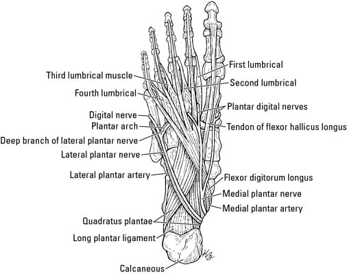
The lateral plantar nerve (external plantar nerve) is a branch of the tibial nerve, in turn a branch of the sciatic nerve and supplies the skin of the fifth toe and lateral half of the fourth, as well as most of the deep muscles, its distribution being similar to that of the ulnar nerve in the hand.. It passes obliquely forward with the lateral plantar artery to the lateral side of the foot ...

Anatomy Of The Lateral Plantar Nerve Everything You Need To Know Dr Nabil Ebraheim Youtube
The plantar nerves are a pair of nerves innervating the sole of the foot. They arise from the posterior branch of the tibial nerve.

Nerves Of The Foot Foot Ankle Orthobullets
04/11/2020 · Foot Pain Diagram. Written By: Chloe Wilson BSc(Hons) Physiotherapy Reviewed By: FPE Medical Review Board A foot pain diagram is a great tool to help you work out what is causing your ankle and foot pain. There are a whole range of structures e.g. bones, muscles, tendons and nerves which will each give slightly different foot pain symptoms.

Healthcrib The Foot Nerves Of The Foot That Distribute Information Via The Sensory Pathway Somatic Nervous System Back To The Central Nervous System Receive A Response Via The Motor Pathway Allowing
Dermatomes exist for each of these spinal nerves, except the first cervical spinal nerve. Sensory information from a specific dermatome is transmitted by the sensory nerve fibers to the spinal nerve of a specific segment of the spinal cord. The C1-C7 nerve roots emerge above their respective vertebrae; the C8 nerve root
Plantar Nerve Wikipedia
The thinnest part of your foot, usually found towards its center, is known as the waistline. Parts of your foot correlated with the stomach are found above the waistline. Parts correlated with the intestines are found below. The bottom of your foot is connected with your pelvic area. Even the basics can lead you to a very simple self-treatment.
Topographical Anatomy Of The Foot And Ankle Lateral Aspect And Nerves
The #posterior leg #muscles that insert on the foot are the: gastrocnemius, plantaris, soleus, tibialis posterior, flexor digitorum longus, and flexor hallucis longus. Collectively, the posterior leg muscles work to plantarflex and invert the foot. They are innervated by the tibial nerve. #foot_muscles #gastrocnemius #flexor_digitorum_longus
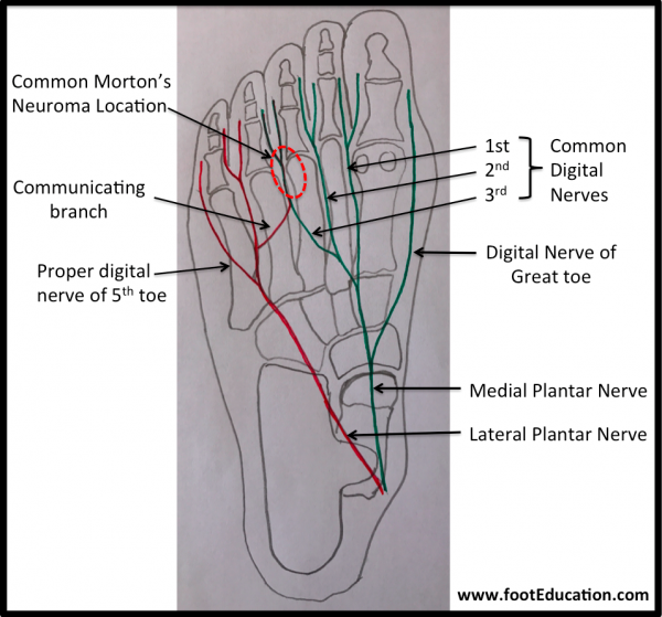
Morton S Neuroma Footeducation
13/04/2015 · They are themselves branches of the larger intermediate dorsal cutaneous nerve, medial dorsal cutaneous nerve, sural nerve, and deep fibular nerve in the lower extremities. Last medically reviewed ...

A Patient S Guide To Foot Anatomy 2020 Orthonorcal Los Gatos Capitola Morgan Hill Watsonville Ca
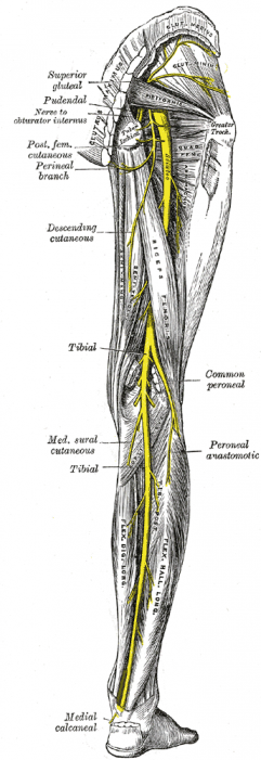
01/01/2019 · The sciatic nerve is about the size of the thumb, quite large as nerves go. The sciatic nerve runs along the gluteus maximus and down the rear portion of the leg. The sciatic nerve splits into several branches past this point and continues traveling down into the foot.

Nerves Of The Foot Clipart Etc Anatomy Organs Human Anatomy Chart Ankle Anatomy
3%. (69/2724) 3. It is the terminal branch of the superficial peroneal nerve; injury leads to reduced sensation over medial aspect of great toe. 83%. (2254/2724) 4. It is the terminal branch of the deep peroneal nerve; injury leads to first interphylangeal joint flexion weakness. 3%.

Welcome To Netter Images
Sural Nerve. The fourth nerve of the foot is another branch of the tibial nerve, known as the sural nerve (Figure 17). This nerve runs from slightly below the knee to the lateral aspect of the foot. It becomes a very superficial nerve at the level of the posterolateral ankle and continues distally to provide sensation to the outside of the foot.
What Is The Anatomy Of The Tibial Nerve Relevant To A Posterior Tibial Nerve Block
Dr. Ebraheim's educational animated video describes the nerves of the lower leg in a very easy and simple animation.Lateral cutaneous nerve of the calfSural ...
Vector Illustration Diagram Nerves Cutaneous Innervation Stock Vector Royalty Free 513767140
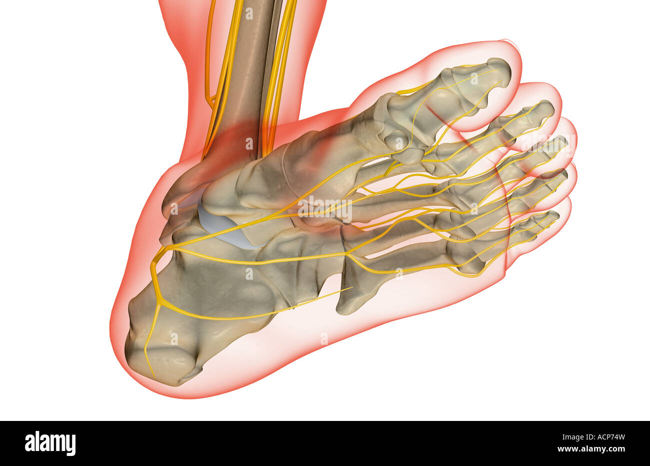
The deep peroneal nerve branches off from the common fibular nerve at the top of the back of the lower leg on the side of the fibula bone. Fewer nerves of the foot are located on the bottom surface than on the top of the foot, even though this is the surface of the foot that comes in contact with the ground.

The Leg Ankle And Foot Knowledge Amboss
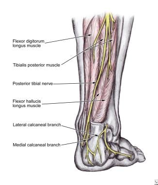
Tibial nerve: This nerve is a branch of the sciatic nerve. It runs down the leg, between the heads of the gastrocnemius, and passes under the soleus. It curves under the medial malleolus and continues into the foot. It innervates all the muscles in the posterior compartment of the leg. Common fibular (peroneal) nerve: This nerve branches off ...
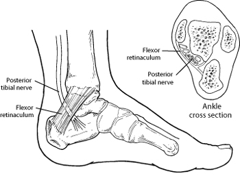
Tarsal Tunnel Syndrome Symptoms Of Tarsal Tunnel Syndrome Foot Health Facts Foot Health Facts
Sciatic nerve. The sciatic nerve is the dominant nerve that innervates the lower back and the lower extremities. It travels from the lower spine, through the pelvis, and down each leg. It is the ...

Foot Nerves Images Stock Photos Vectors Shutterstock
The nerves of the foot help move the body and keep balance both while it's moving and at rest. All of these nerves extend as branches of nerves in the leg that pass through the ankle and into the foot. The sural nerve branches from the tibial and common fibular nerves and is responsible for feeling on the outside of the foot and the small toe.

A Patient S Guide To Foot Anatomy 2020 Orthonorcal Los Gatos Capitola Morgan Hill Watsonville Ca
The recurrent laryngeal nerve (RLN) is a branch of the vagus nerve (cranial nerve X) that supplies all the intrinsic muscles of the larynx, with the exception of the cricothyroid muscles.There are two recurrent laryngeal nerves, right and left. The right and left nerves are not symmetrical, with the left nerve looping under the aortic arch, and the right nerve looping …

Cutaneous Nerve Supply Of The Foot Diagram Quizlet
This image is titled nerves of the leg diagram and is attached to our article about Leg Nerves and Reflex Motion in Feet.. Be sure to visit the guide for more context and information about nerves of the leg diagram, or read some of our other Health & Anatomy posts!

Medial And Lateral Plantar Nerve Entrapment Musculoskeletal And Connective Tissue Disorders Merck Manuals Professional Edition
Problems with nerves in the feet are very common. Many times, an injured nerve will cause intense pain and heat felt within the foot. Nerves act as a network, communicating important information from the foot to the brain. Learn more about the various conditions and problems that can affect the nerves in the foot.

Ankle Block Landmarks And Nerve Stimulator Technique Nysora
Nerve pain in foot can stem from two places. The Spine: Damage to the lower back is a common cause of nerve pain in the foot. This tends to cause back, buttock and leg nerve pain as well as foot pain and weakness. The Nerve: a peripheral foot neuropathy is caused by damage along the course of the nerve somewhere down the leg or in the foot itself.
Lower Limb Arteries And Nerves Anatomy Branches Kenhub
normal anatomy and common variants of the nerves of the foot and ankle, with use of dissected specimens and correlative US and MR imaging findings. In addition, the authors illustrate proper probe positioning, which is essential for visualizing the nerves at US. The authors' discussion focuses on the superficial and deep

Sciatic Nerve Anatomy
1

The Diagnostic Anatomy Of The Sciatic Nerve Neupsy Key
Common Peroneal Nerve Physiopedia
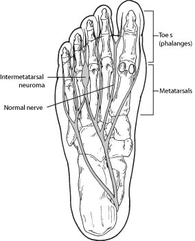
Morton S Neuroma Symptoms Of Morton S Neuroma Foot Health Facts Foot Health Facts

Compression Neuropathy Lincoln Park Lakeview Chicago Il Lincoln Park Podiatry
Nerves In The Foot Dummies

Normal Anatomy And Compression Areas Of Nerves Of The Foot And Ankle Us And Mr Imaging With Anatomic Correlation Radiographics
The Nerves Of The Foot Stock Photo Alamy
Foot And Ankle Anatomy Video Foot Ankle
1
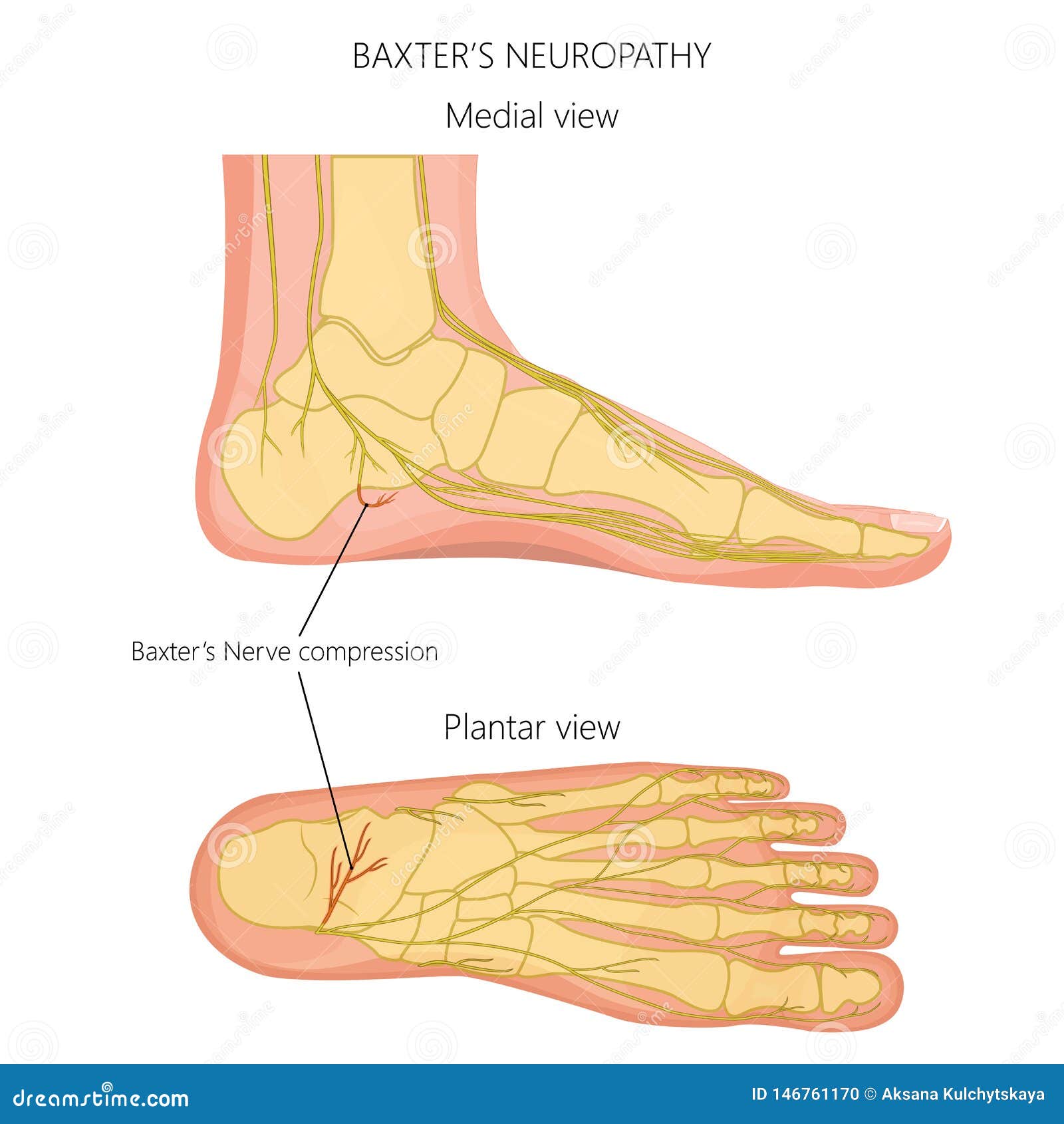
Nerves Of The Foot 02 Stock Vector Illustration Of Mortons 146761170

The Leg Ankle And Foot Knowledge Amboss

The Tibial Nerve Course Motor Sensory Teachmeanatomy
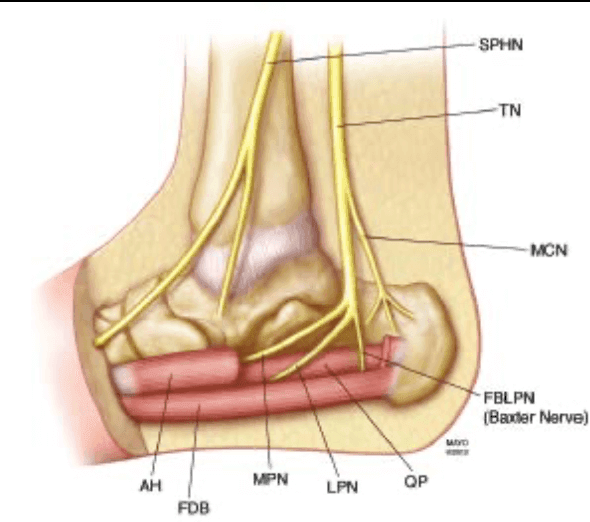
Baxter S Nerve Entrapment Ultrasound Guided Injections

Layers Of The Plantar Foot Foot Ankle Orthobullets


Comments
Post a Comment