38 clam diagram labeled
two bamboo skewers. · clam diagrams. Procedures: 1. Teacher prepares clams ahead of time by placing in boiling water until the adductor muscles relax. (DO. Removal of the mantle shows the underlying soft body parts, a prominent feature of which are the adductor muscles in dimyarian species (clams and mussels) or ...
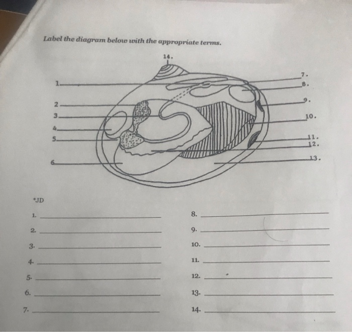
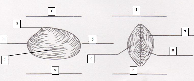
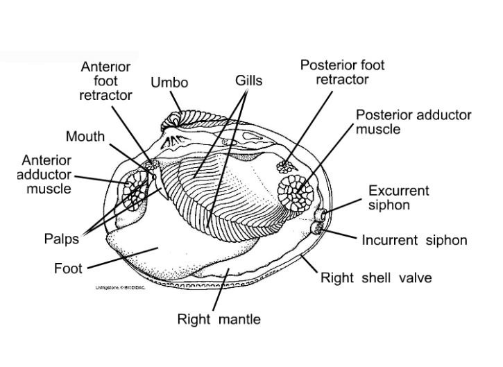
Begin to identify and label the external anatomy and their functions on your worksheet or in your lab notebook. 4. Follow the teacher's instructions and your ...
Clam diagram labeled
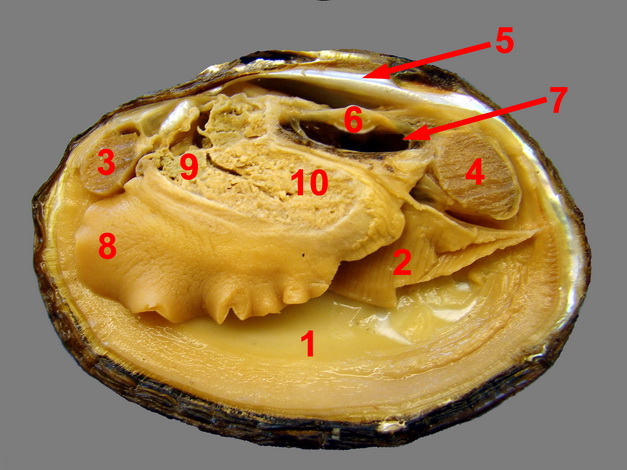
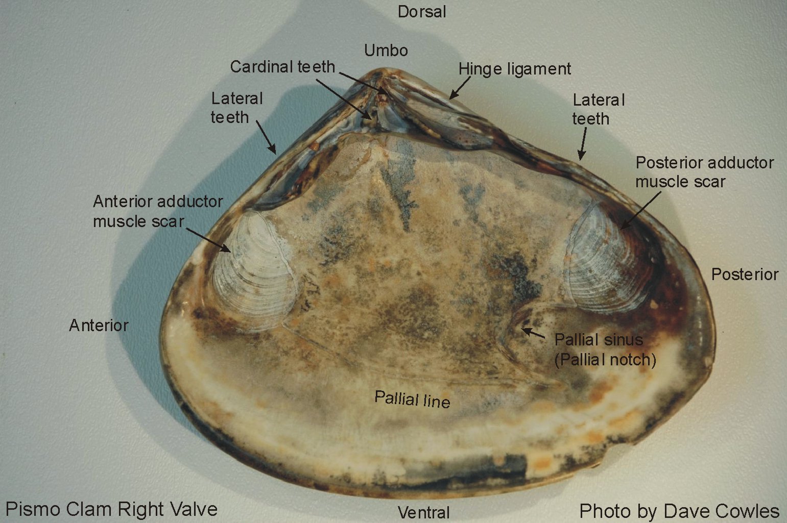
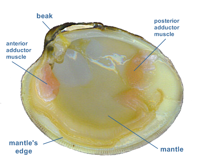
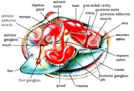
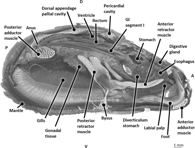
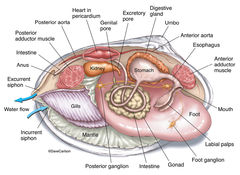
They do this by siphoning water over the gills which traps it and propels it toward the mouth. Related Links. Coloring Page · Labeling Page Based on histologic research in the giant clam Tridacna gigas, ... General anatomy of the blue mussel (Mytilus edulis) tissues. Find the anus just behind the posterior adductor muscle. 21. Draw a picture of what you see and label the following structures: Umbo. Adductor Muscles.
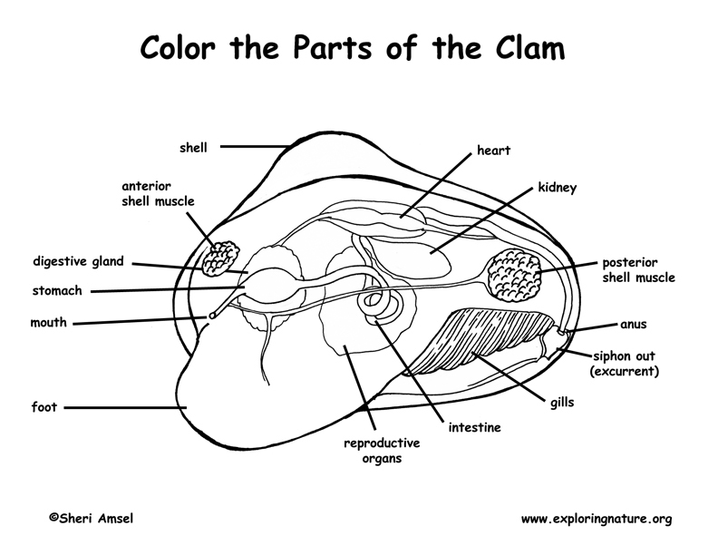
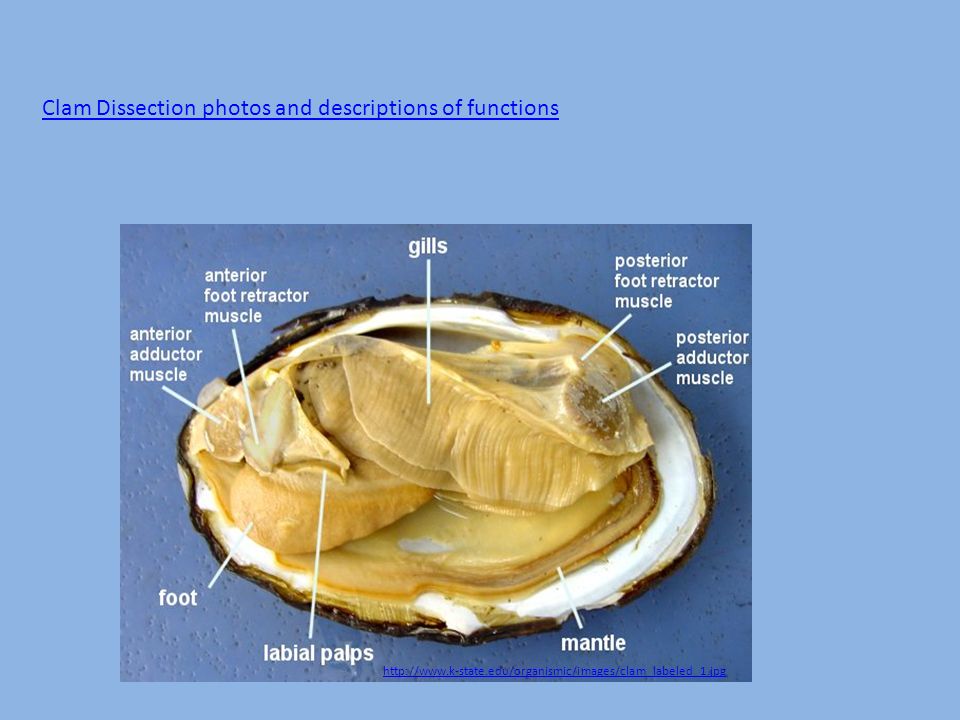
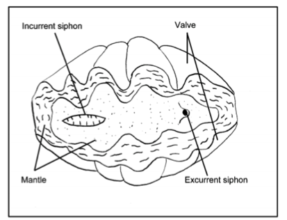
Clam diagram labeled. Learn the external and internal anatomy of the clam and squid ... Most are familiar to you as food sources: oysters, clams, scallops, and yes, snails, squid. Label the Parts of the Clam Science Lessons, Science Activities, Nervous System Anatomy,. sheriamsel. Exploring Nature Eduational Resource. 10k followers. To study the internal and external anatomy of a bivalve mollusk. ... on your lab report & label the diagrams of the internal structures of the clam. Image result for clam anatomy. BACKGROUND: Clams are bivalves, ... B. Using the words in the above table label the following diagrams of the clam.
Find the anus just behind the posterior adductor muscle. 21. Draw a picture of what you see and label the following structures: Umbo. Adductor Muscles. Based on histologic research in the giant clam Tridacna gigas, ... General anatomy of the blue mussel (Mytilus edulis) tissues. They do this by siphoning water over the gills which traps it and propels it toward the mouth. Related Links. Coloring Page · Labeling Page
2
2

Untitled Page

Clam Dissection Docx
Lab 6 Molluscs Zoo Lab Uw La Crosse
Clam Dissection Biology Junction
Review Guide Mollusks And Annelids
Glossary
Hard Clams Barnegat Bay

Tridacna Squamosa Fluted Giant Clam Taxo4254 Wiki Nus

The Hatchery Culture Of Bivalves A Practical Manual
Solved Class Bivalvia Clams Scallops Ovsters And Mussels Chegg Com
Anatomy Of Animals

Clam Anatomy Flashcards Quizlet

Bivalve Clam Diagram Quiz

Clam Dissection Lab Explained Schoolworkhelper
33 Label The Internal Structures Of The Clam Labels Design Ideas 2020
The Blue Mussel Inside 3d Visualization And Description Of The Vascular Related Anatomy Of Mytilus Edulis To Unravel Hemolymph Extraction Scientific Reports
Untitled 1
Clam Anatomy Coloring Page
Mollusk Students Britannica Kids Homework Help

The Hatchery Culture Of Bivalves A Practical Manual
Clam Dissection Diagram Quizlet

New Page 1

Clam Dissection Docx

Clam Anatomy101 Clam Anatomy Diagram Tanks4thememories Flickr
Mollusks Clam Dissection Bivalve Information Ppt Download
Clam Dissection Introduction
2
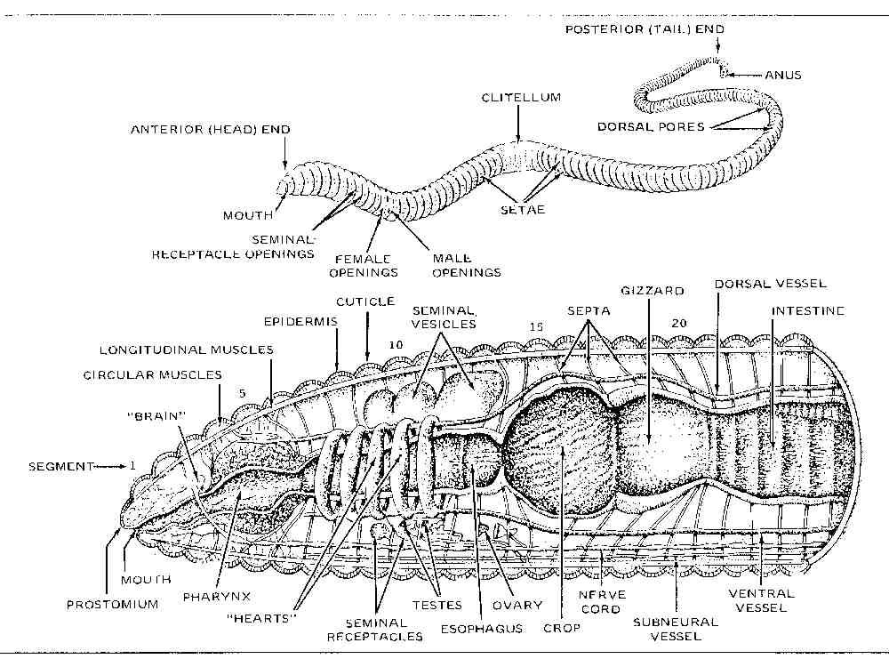
Worm Biology 101
2

Giant Clams Clamsplaining

An Overview Of The Most Important Terminology Needed For Orientation Of Download Scientific Diagram
Zoology Carlson Stock Art

Images For Bio 122 Lab
Clam Visceral Mass
Taxo4254 Tridacna Squamosa

Basic Clam Anatomy Internal Quiz


Comments
Post a Comment