43 mink muscle diagram
Since these mink box plans are pretty basic and only require a few common tools, the project will only take you about 30 minutes or less to complete. Although you can use scrap plywood, I like to use a piece of 1'' x 8'' pine board. The boards actually measure .75'' x 7.25'' making them a great width for the mink box. Muscle Diagrams Labels Nov 6, Can you name the Mink Muscles? Test your knowledge on this science quiz to see how you do and compare your score to others. Quiz by. A Guide to the Dissection of the Mink .. 5 Lateral view of the Mink Skull (drawn from specimen). 33 Muscles of the Eye (diagrammatic representation).
Mink Dissection Guide Muscles Dissection Muscle Diagram Muscle . Is There Proof God Exists Human Body Systems Body Systems Body Cells . Pin Em Medicina Veterinaria . Free Art Print Of Diagram Of Human Bone Anatomy Human Bones Anatomy Human Bones Human Anatomy And Physiology .
Mink muscle diagram
body of mandible below incisors skin & muscle @ angle of mouth (below insertion of zygomaticus) • draws corner of mouth laterally & downward • antagonist of zygomati-cus Facial Depressor labii inferioris 17 body and mandible lateral to its midline skin & muscle of lower lip • draws lower lip inferiorly (pout) Facial Orbicularis oris 18 Mink Muscle Dissection Head, Neck, and Shoulder - YouTube 17 Lateral Muscles ............. 18 Ventral View of Viscera ..... 19 Dorsal View of Pharyngeal Region - Tongue Cut and Hyoid Attached (drawn from specimen). 20 ...32 pages
Mink muscle diagram. Badger Body Diagrams and Habitat Posters. Bandicoot (Bilby) Body Diagrams and Habitat Posters. Bat (Little Brown) Body Diagrams and Habitat Posters. Bat (Townsend's Big-eared) Diagram. Bear (Black) Body Diagrams and Habitat Posters. Bear (Grizzly) Body Diagrams and Habitat Posters. Bear (Polar) Body Diagrams and Habitat Posters. flexes and rotates vertebral column. external abdominal oblique. compresses abdominal wall, laterally rotates trunk. pectoralis major. white; Adducts and flexes humerus. pectoralis minor. blue; protracts and depresses scapula. Trapezius. Elevates, depresses, retracts, and rotates the scapula; ... Mink Muscle Labeled Diagram. August 30th, 2013 19:46:27 PM . ART 1: CLASSROOM RULES - SchoolWorld an Edline Solution D Make sure the outer bag is labeled with your names and hour. ... The heart muscle capillaries are drained by a ... Mink Dissection Activity #1 page 9 Diagram 7: ... [Filename: ... Start studying Mink Muscle Anatomy (G&M). Learn vocabulary, terms, and more with flashcards, games, and other study tools.
Study 13 mink muscle anatomy flashcards from Shelly O. on StudyBlue. Telltale signs of a mink kill are usually signs of a fierce struggle and the bird has injuries to the back of its head and neck or may have its head removed. Taking a closer look at the injury, you are likely to see the marks from the mink's pairs of closely spaced canine teeth. Mink usually do not eat the whole animal the first night. The whole skull anchors muscles that hold the head up and allows the mink to chew. Most of the bones of the skull are flat bones, except the mandible. The mandible is attached to the skull by a type of hinge joint. It is the biggest, strongest bone of the skull. The dental formula of the mink is: 3/3, 1/1, 3/3, 1/2 = 34 teeth. Start studying Mink Muscles. Learn vocabulary, terms, and more with flashcards, games, and other study tools.
Female Muscles In The Body Diagram : Mink Internal Organs Part 2.wmv - YouTube / The major muscle in the chest is the pectoralis major. October 19, 2021 When the male body is the default in biology classes, medical education . These muscles are also in a woman's uterus, which is where a baby develops. The laminated, color female muscular system chart is extensively detailed and ideal for ... 3 Sept 2015 — Advanced Biology Human Anatomy Mink Dissection of Muscles In this portion of our study of the Mink, we will be focusing on the muscular ... Mink Muscle Dissection. You need gloves Mink Muscle Diagrams. Mink Muscle Dissection. Mink Muscle Web Site Images - no labels. Comments. Sign in | Recent Site Activity | Report Abuse | Print Page | Powered By Google Sites ... Sign In. You may be offline or with limited connectivity. Try downloading instead.
TODAY'S AGENDA DISSECTION TOOLS
Practice Practical Identify these muscles Close up Answer 1. Rectus Femoris 2. Gracilis Identify this muscle Answer 3. Clavotravezius Identify this muscle Answer 4. Serratus ventralis (Serratus anterior in humans) a. Identify this part of the anatomy. b. Which way does the blood flow?

Mink Skull Diagram and Labeling
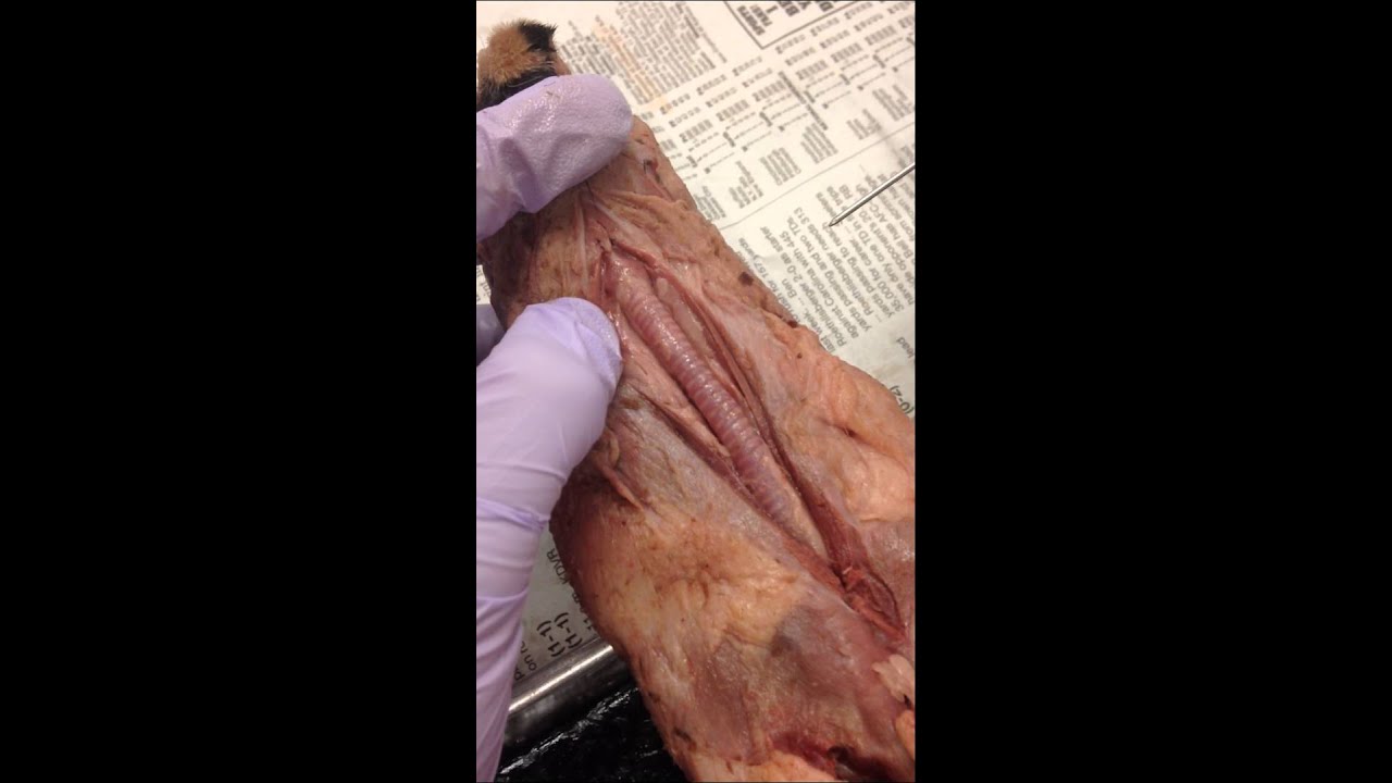
Lab 3: Mink back & Shoulder Muscular System (Smith & Schenk, A Dissection Guide and Atlas to the Mink, 2000) Muscles are designed with one basic purpose in mind - movement. Muscles work to either move an animal through its environment or move substances through an animal. In vertebrates, there are three basic types of muscle tissue - skeletal muscle and cardiac muscle, both of which possess ...

Animal Lab - Mink/Cat Musculature Flashcards | Quizlet
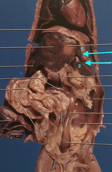
The circulatory system of both, the humans and the mink, begin at the heart. The heart is virtually in the middle of the chest with all of the arteries and veins attached at the base. A unique characteristic of the heart is that it is an involuntary muscle. It is a muscle that we cannot control based upon our choosing, unlike skeletal muscles.

Mink Anatomy
May 2, 2017 - Play this quiz called Mink Muscles Identification and show off your skills.
Mink Dissection by Brittany Jerlinga | Teachers Pay Teachers
Oak Park Unified School District is making improvements to all of its campuses in innovative ways. We would like to thank our community for its support in passing Measure S and allowing us to provide a modern and safe learning environment for our students · The Student Nutrition & Wellness ...
Mink Abdominal Cavity Flashcards | Easy Notecards
Mink Dissection of Muscles In this portion of our study of the Mink, we will be focusing on the muscular system. These instructions tell you what muscles you are to identify and provide instructions on information you are expected to learn about those muscles. Additionally, these instructions will reference passages in the
Muscular System - Mr. Smit: Life Sciences For SHS
Mink muscles arm 2310.key Author: Anil Rao Created Date: 10/24/2017 8:19:59 PM ...

Heritage Associates LLC
Mink Dissection Labeled Diagrams. TODAY'S AGENDA. 1. Dissection Basics & Clean-up. 2. Remove external fat from mink. 3. Locate muscles of the neck and. labeled #1. What is the common name for this muscle? Presentation on theme: "Mink Dissection Practice Practical. .. Muscle Diagrams Labels Anatomy and Physiology.

Mink - Muscles - YouTube
muscle Tongue papillae Ear canal Hard palate roof of mouth. Digestive system-parotid duct Parotid salivary gland Parotid duct Masseter muscle. Esophagus Digestive system- esophagus Probe under trachea and esophagus Trachea. ... Digestive system mink.key Author: Anil Rao Created Date:
PPT - Superficial Muscles of the Body - Anterior View ...
Note : Vastus intermedialis is deep to the rectus femoris. Vastus medialis. Vastus lateralis. Rectus femoris*. Sartorius reflected. Sartorius reflected ...10 pages

Pinterest • The world's catalog of ideas
Mink Veins Diagram. Upper Body - (Cat manual) Lower Body - (Cat manual) Pig Heart Dissection (Instructions) Pig Heart Dissection (Questions) Quiz Materials. Vein Quiz Diagram; Artery Quiz Diagram ; Heart Quiz Diagram; Essays to get out of a section of the Circulatory test . Notes & Diagrams. Circulatory notes; Blood Vessel Diagrams - Blank
MINK DISSECTION
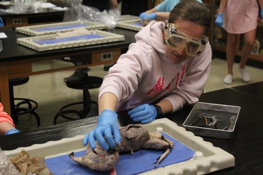
THE MUSCULAR SYSTEM. Your mink has been skinned for you. Your task now is to clean the fat and fascia off so that muscle fiber directions can be seen and to separate each muscle from adjacent ones. Fingernails and the blunt probe are most useful. When cutting a muscle, first separate it completely ...
Mink
1) This Checklist is what you will be turning in. You will need to be able to actually identify these structures during the practical. 2) This Handout will be given to you to use during lab. 3) "The Sets" are pictures referenced in the handout. You will need these during lab and can access them online. Č.

Mink Dissection by Brittany Jerlinga | Teachers Pay Teachers
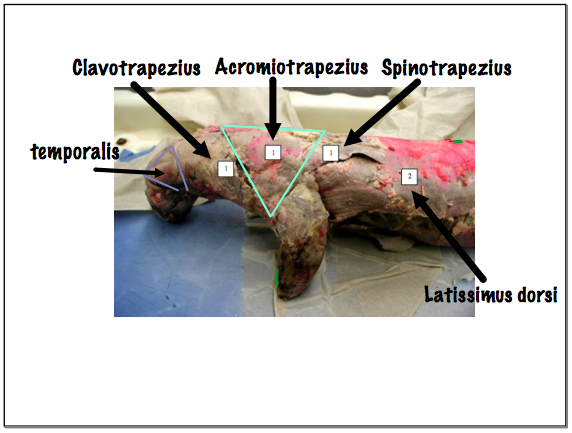
Mink Dissection 1. Mink dissection Hi! 2. Platysma, Panniculus Carnosum, Facia Facia platysma I'm guessing that's the panniculus carnosum because it's the "bacon" and on that side too the white parts are platysma 3. Latissimus Dorsi The whole side of the mink is the latissimus dorsi 4.

Mink Dissection #1 St. Brendan High School 2012 - YouTube
Anatomy & Physiology. Mink Muscles. Thorax – Ventral. 1. Masseter. 2. Sternomastoid. 3. Pectoralis Major. 4. Pectoralis Minor. 5. External oblique.1 page
Mink Dissection
Cat muscular diagram and labelling practice; Mink Muscular system (PDF) Mink Dissection of Muscles (PDF) Mink Video dissection of Head, Neck, and Shoulder; Mink Video dissection of Abdomen, Limb, Hind limb
Mink Dissection of Muscles - highschoolbiologywiki Pages 1 - 13 - Flip PDF Download | FlipHTML5
The pectoral region is located on the anterior chest wall. It contains four muscles that exert a force on the upper limb: the pectoralis major, pectoralis minor, serratus anterior and subclavius. In this article, we shall look at the anatomy of the muscles of the pectoral region - their attachments, actions and innervation.
Lab Objectives, BIO 2310, Spring 2018 | Clare Hays Biology Homepage
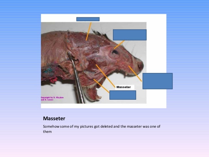
Mink muscle diagram. Locate as many of them as you can on the diagrams below. About press copyright contact us creators advertise developers terms privacy policy safety how youtube works test new features press copyright contact us creators. Sign in recent site activity report abuse print page powered by google sites. Start studying mink muscles. Mink dissection practice practical. Mink muscle ...
Mink Dissection 1: The Throat Region - YouTube
the individual muscles. However,using scissors or scalpels may result in cutting muscles or other structures. Blunt dis-section is a technique that uses blunt probes and forceps to remove fascia and separate muscles. To observe a deep muscle,you will have to cut the superficial muscle at the midline and reflect (pull back) the edges toward the ...

Anatomy of rat musculature, ventral view. | Aleatória
The scalenus anterior muscle is the anteriormost of the three scalene muscles. It originates from the anterior tubercles of transverse processes of the vertebrae C3-C6. The muscle takes an inferior, almost vertical, course towards the thoracic cage.It gives off a single flat tendon, that inserts onto the scalene tubercle and superior border of first rib, just anterior to the groove for ...

Mink Muscle Dissection Head, Neck, and Shoulder - YouTube
Cub Cadet 1180 Series 1000 Model 40777 Wiring Diagram. 04.09.2018 04.09.2018. 1263 1264 1265 1266 1267 1268 1269 1270 1271 1272 1273
Neck Muscles 1. Digastric 2. Mylohyoid 3. Geniohyoid 4. Sternohyoid ppt video online download
Human Muscles Labeled Diagram For Kids Infographics Muscle. C Ous Atae M Figure 311 Illustrates The Ven Cheggcom. Royalty Free Stock Illustration Of Diagram Dorsal Ventral Cavities. A Motor And A Brake Two Leg Extensor Muscles Acting At The Same. Muscle Diagrams To Label Diagram Dorsal Cat Oasissolutionsco.
TODAY'S AGENDA DISSECTION TOOLS
17 Lateral Muscles ............. 18 Ventral View of Viscera ..... 19 Dorsal View of Pharyngeal Region - Tongue Cut and Hyoid Attached (drawn from specimen). 20 ...32 pages

Frog Sculpture, Dunston, Gateshead, Tyne & Wear, England.
Mink Muscle Dissection Head, Neck, and Shoulder - YouTube

Ferret (Ictis) | Flickr - Photo Sharing!
body of mandible below incisors skin & muscle @ angle of mouth (below insertion of zygomaticus) • draws corner of mouth laterally & downward • antagonist of zygomati-cus Facial Depressor labii inferioris 17 body and mandible lateral to its midline skin & muscle of lower lip • draws lower lip inferiorly (pout) Facial Orbicularis oris 18
Mink Dissection Practice Practical. - ppt video online download

Mink Dissection: Neck and Back Muscles - YouTube
Human Anatomy- Mink Muscles (Shoulder Muscles) Flashcards | Quizlet

Untitled Document | Cat anatomy, Animal science, Human ...

1000+ images about Cat muscles on Pinterest | Muscle, Cats ...
Practical 3 Flashcards | Chegg.com
Mink Anatomy

Cat dissection lab_labeled_images

Mink Anatomy

1000+ images about Anatomy on Pinterest | Human anatomy ...

Mink Muscle Dissection Preview Group 5 - YouTube

PPT - Sample Mink Dissection Quiz PowerPoint Presentation ...
26 Cat dissection muscles ideas | cat anatomy, dissection, anatomy

Mink Dissection - YouTube

Mink Anatomy

annotated transfer case diagram | JEEPS | Pinterest | Jeep ...

22 best images about Anatomy..Creating a New Aspect. on ...
Pronator Teres Muscle Origin, Function & Anatomy | Body Maps


Comments
Post a Comment