39 replication fork diagram labeled
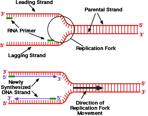
October 3, 1996 - In this diagram of the process of DNA replication at a replication fork, the black boxes labeled D and E are: The replication fork moves at the rate of 1000 nucleotides per second. DNA polymerase can only extend in the 5′ to 3′ direction, which poses a slight problem at the replication fork. As we know, the DNA double helix is anti-parallel; that is, one strand is in the 5′ to 3′ direction and the other is oriented in the 3′ to 5′ direction.
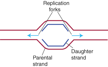
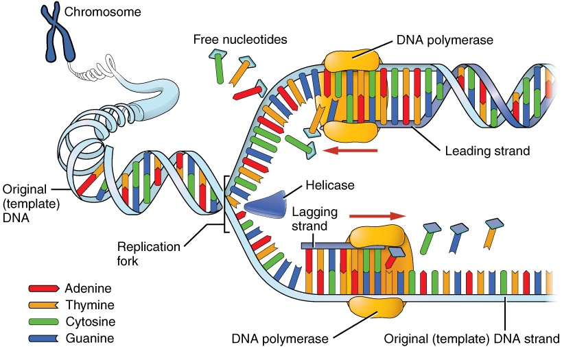
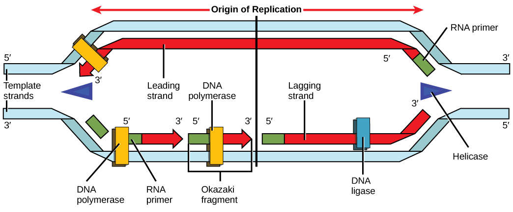
replication begins at an origin of replication where two strands are separated opening a replication bubble. A eukaryotic chromosome may have hundreds or thousands of origins of replication and proceeds in both directions from origin. At the end of each bubble is a replication fork.

Replication fork diagram labeled
replication fork: the Y-shaped structure formed during the initiation of replication semiconservative replication: the method used to replicate DNA in which the double-stranded molecule is separated and each strand acts as a template for a new strand to be synthesized, so the resulting DNA molecules are composed of one new strand of nucleotides ... November 1, 2016 - DNA replication has been well studied in bacteria primarily because of the small size of the genome and the mutants that are available. E. coli has 4.6 ... 21 Jul 2021 — One of the strands is oriented in the 3' to 5' direction (towards the replication fork), this is the leading strand?. The other strand is ...
Replication fork diagram labeled. In this diagram of the process of DNA replication at a replication fork, the strand labeled B is the: DNA Replication. The two antiparallel strands are replicated simultaneously in both directions. RNA primers are used to initiate a new strand. The advancing replication forks meet at a point opposite to the point of origin thus opening up the coiled DNA molecule. This is called θ (theta) model of replication. But the unwinding is a very complex mechanism as the two strands are coiled. The two advancing replication forks make the remaining entire un-replicated portion of DNA overwound. The replication fork. In this diagram of the process of DNA replication at a replication fork, the strand labeled B is the: . The replication fork is a very active area where DNA replication takes place. It is created when DNA helicase unwinds the double helix structure of the DNA. The replication fork looks like a fork ...
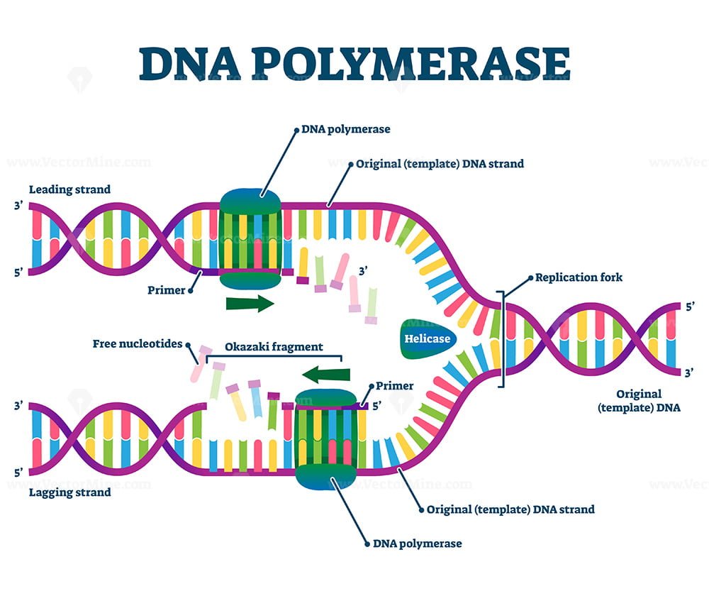
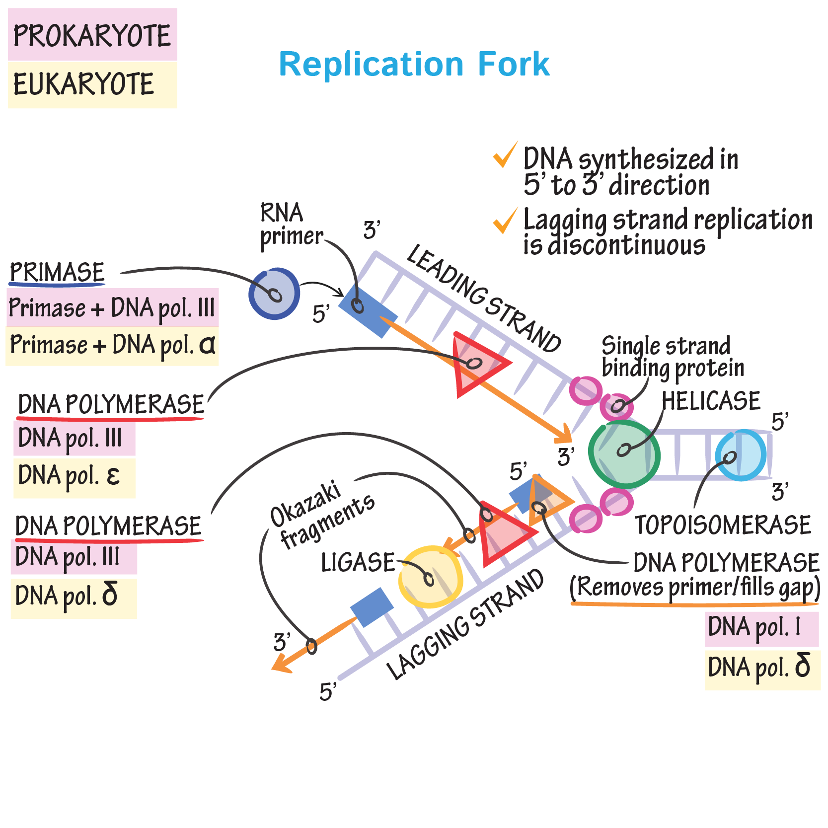
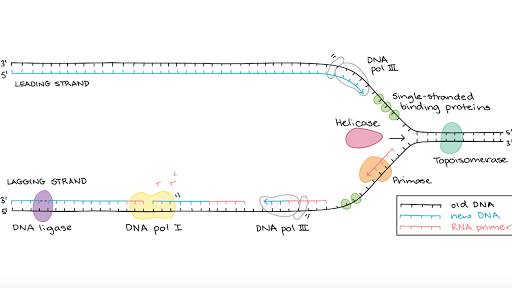
Click here to get an answer to your question ✍️ Draw a labelled diagram of replicating fork.1 answer · Top answer: Labelled diagram of replication fork: Dna replication diagram labeled. Elongation is the second stage, in which one strand of dna or the template strand works as a template for rna polymerase. Label the diagram below with the following choices. The density gradient centrifugation permit very small differences like between 15n and 14n labeled dna to be detected. DNA ligase joins the Okazaki fragments together into a single DNA molecule. Helicase opens up the DNA at the replication fork. Single-strand binding proteins coat the DNA around the replication fork to prevent rewinding of the DNA. Topoisomerase works at the region ahead of the replication fork to prevent supercoiling. The strand, which is synthesized in the same direction as the replication fork, is known as the 'leading' strand. The template for this strand runs in the direction of 3' to 5'. The Polymerase has to attach only once and it can continue its work as the replication fork moves forward.
January 29, 2013 - The replication fork. Leading-strand synthesis proceeds continuously in the 5' to 3' direction. Lagging-strand synthesis also occurs in the 5' to 3' direction, but in a discontinuous manner. An RNA/DNA primer (labeled in green) initiates leading-strand synthesis and every Okazaki fragment on ... Working with Molecular Genetics Chapter 5, DNA Replication I, v2 3 shifted to grow in media containing the highly abundant, light isotope of nitrogen, 14N, in the NH4Cl, so that newly synthesized DNA would have a "light" density.The labeled, heavy (old) Dna Replication Diagram Labeled. dna replication with diagram the other two were hybrid molecules hl this proves that during replication one parent strand is conserved and the other new strand is synthesized thus dna replication is a semi conservative process enzymes of dna replication the enzymes which take part in replication are able to copy dna molecules which may contain millions of bases ... DNA Replication Definition: For the growth of an individual, cell division is a necessary part. When the act of cell division occurs, the DNA must be replicated. During cell division, the DNA successfully copied in the daughter cells. Many enzymes take place for this act. The DNA has to be inherited and copied in two daughter cells.
2 511 Dna Replication Stock Photos Pictures Royalty Free Images Istock
In The Replication Fork Below Label The L Clutch Prep . Discontinuous Dna Synthesis Composed Of Okazaki Fragments. Dna Replication Labeling Genetics Quiz Quizizz : It Is The Process By Which An Organism's Dna Is Replicated To In The Diagram Above, The Strand On The Right Would Be The Leading Strand.
Can Anyone Help A Girl Out With Labeling A Replication Fork Diagram 4 Of Them Are Correct But It Doesn T Tell Me Which Are Right Wrong R Biology
The replication fork is a region where a cell's DNA double helix has been unwound and separated to create an area where DNA polymerases and the other enzymes involved can use each strand as a template to synthesize a new double helix. An enzyme called a helicase catalyzes strand separation. Once the strands are separated, a group of proteins called helper proteins prevent the
Genes Free Full Text The Replication Fork Understanding The Eukaryotic Replication Machinery And The Challenges To Genome Duplication Html
Written By Pelvic Diagram Sunday, June 7, 2020. Edit. Dna Replication Diagram Labeled. DNA replication occurs through a semiconservative mechanism, because each new molecule is made up of one old strand and one new strand. DNA Replication has three steps - Initiation, Elongation, and Termination. DNA Replication: Steps, Process, Diagram and ...

In The Replication Fork Below Label The L Clutch Prep
ColE1 Replication Control-an example of primer control of replication 1. RNAII will serve as a primer for the replication fork. 2. The 3’ end is processed by host RnaseH to allow efficient RNA-DNA hybrid to form 3. The hybrid acts as a primer for host Pol1 4. As the concentration of plasmid increases, Rop does also 5. Rop stabilizes the RNA1 ...
Sdjohnston Faculty Noctrl Edu
The place where the two strands begin unwinding is called the replication fork. These strands can be used to make new complementary strands called the leading ...

Micro 261 Exam 1 Chapter 4 Mastering Questions Flashcards Quizlet
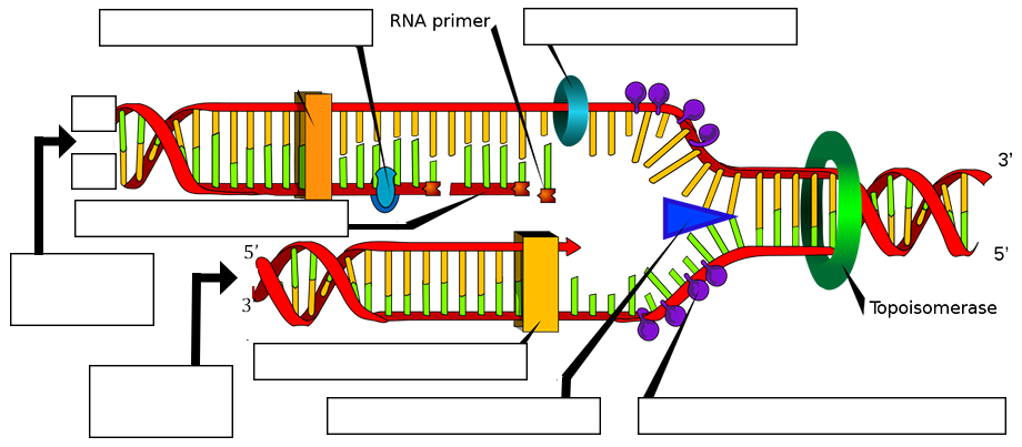
The diagram below shows a replication fork with the two parental DNA strands labeled at their 3' and 5 4/4(5). The diagram below shows a bacterial replication fork and its principal proteins. Drag the labels to their appropriate locations in the diagram to describe the name or function of each structure.
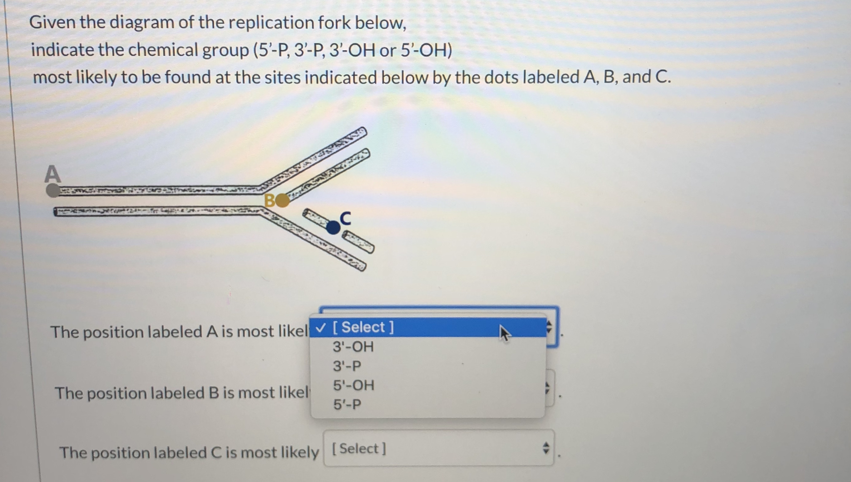
Answered Given The Diagram Of The Replication Bartleby
Figure 1: Replication fork components. The RF is a multiprotein complex with helicase and DNA synthesis activities. It is called a fork because the structure resembles a two-pronged fork. The ...

Questions Label The Image Below Showing A Replication Bubble During Dna Replication According To The Letter Homeworklib
Transcribed image text: The diagram below represents a replication bubble (or fork). The arrows indicate the direction the replication fork is moving, showing how the double stranded DNA is unwinding, А A 5 3 3 B B* 5 2 Replication of the region in the top DNA strand labeled "A" would be a Choose...

Dna Replication Labeling Diagram Quizlet
Below is a replication fork (one side of an origin): a double-stranded DNA ... 5′ ends), leading-strand DNA polymerase and new DNA (label 3′ and 5′ ends and ...7 pages
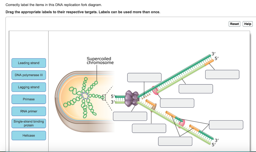
Solved Correctly Label The Items In This Dna Replication Chegg Com
Correctly label the items in this DNA replication fork diagram. Drag the appropriate labels to their respective targets. Labels can be used more than once. Reset | Help Supercoiled chromosome Leading strand DNA polymerase III Lagging strand IIIIIIIIIIIIIIIIIIIIIIIIIIIII Pocong 5' Primase Copcodoro RNA primer Single-strand binding protein Helicase 3

In The Replication Fork Below Label The L Clutch Prep
September 25, 2018 - DNA replication complexes are often prematurely ejected from sites of DNA replication. Left unrepaired, this situation results in incomplete genome replication and cell death. All cells have therefore evolved “replication restart” mechanisms to reload the replicative machinery.
Dna Polymerase Enzyme Syntheses Labeled Educational Vector Illustration Vectormine
June 20, 2018 - Methodologies have been developed to probe the association of proteins with replication forks inside living cells (22, 23). For example, replicating DNA can be pulse labeled by incorporation of a nucleotide analog that allows purification of the labeled DNA together with proteins associated ...

Dna Replication Microbiology
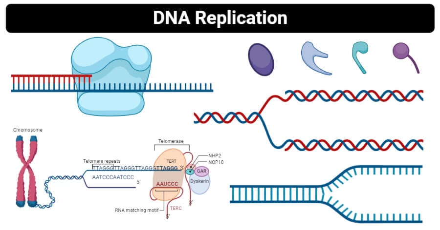
The elucidation of the structure of the double helix by James Watson and Francis Crick in 1953 provided a hint as to how DNA is copied during the process of DNA replication.Separating the strands of the double helix would provide two templates for the synthesis of new complementary strands, but exactly how new DNA molecules were constructed was still unclear.
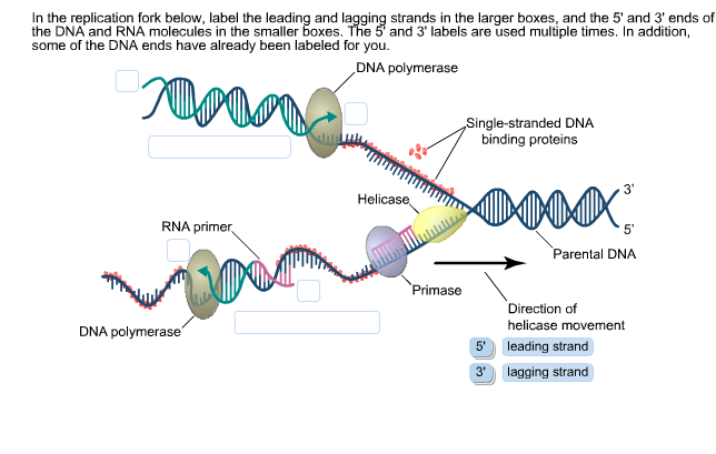
Solved In The Replication Fork Below Label The Leading And Chegg Com
February 25, 2020 - Skip to main page content · The fast and superprocessive KIF1A chemomechanical cycle

Timing Coordination And Rhythm Acrobatics At The Dna Replication Fork Journal Of Biological Chemistry
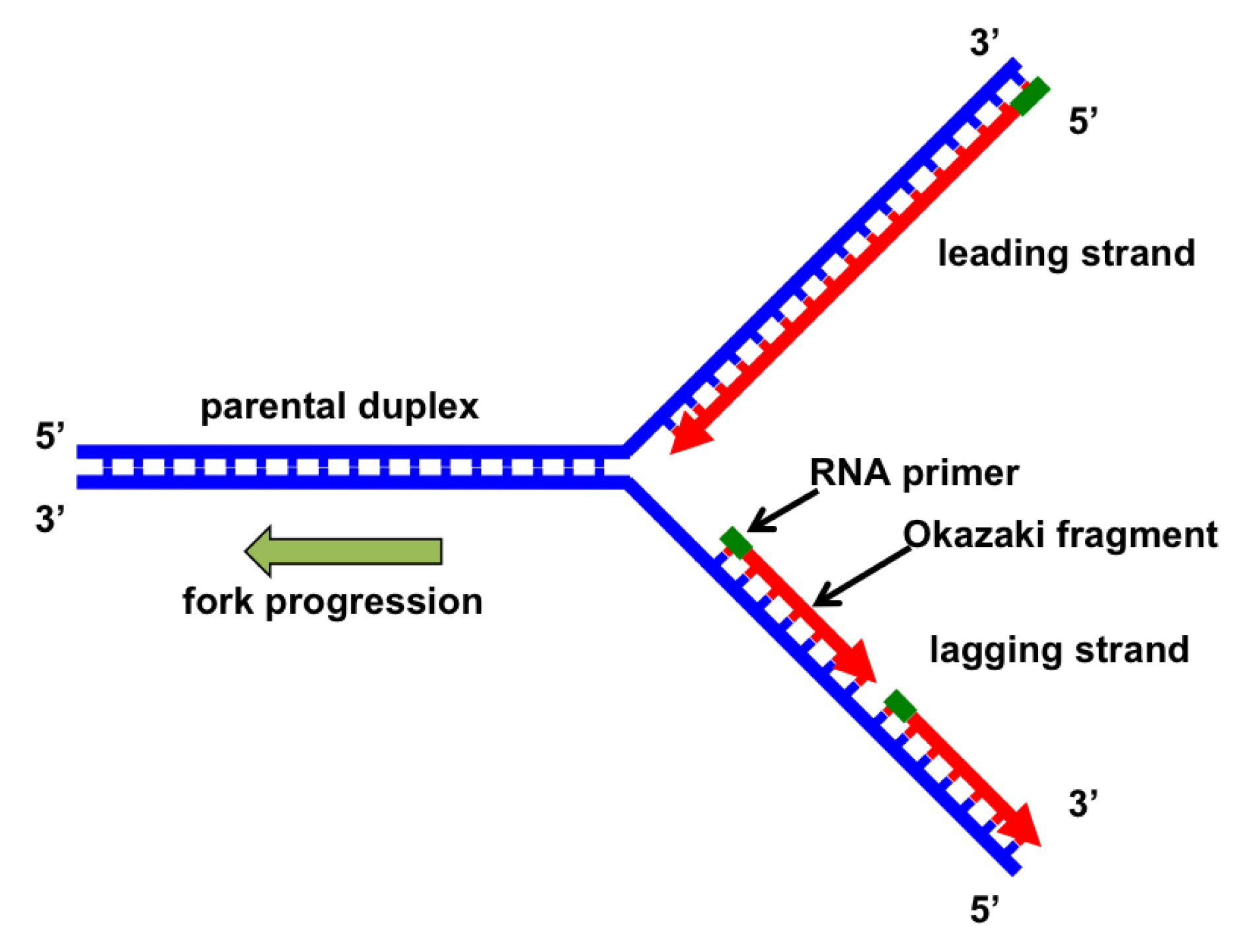
Two replication forks are formed at the origin of replication, allowing for bidirectional replication and formation of a structure that looks like a bubble when viewed with a transmission electron microscope; as a result, this structure is called a replication bubble. The DNA near each replication fork is coated with single-stranded binding ...

34 Label The Diagram Showing Dna Replication Label Design Ideas 2020
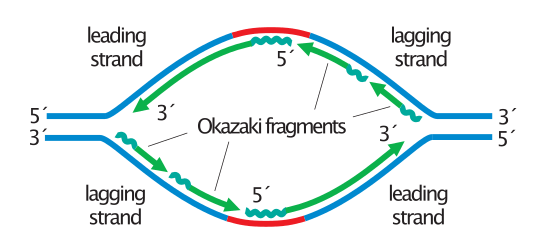
The strand in replication that is copied 3' to 5' as Okazaki fragments and then joined up. DNA Polymerase on lagging strand synthesizes new DNA only in the 3' to 5' direction
Dna Replication Definition Enzymes Steps Mechanism Diagram
Draw a labelled diagram of a “replicating fork” showing the polarity. ... Concept: DNA Replication - The Experimental Proof. Report Error
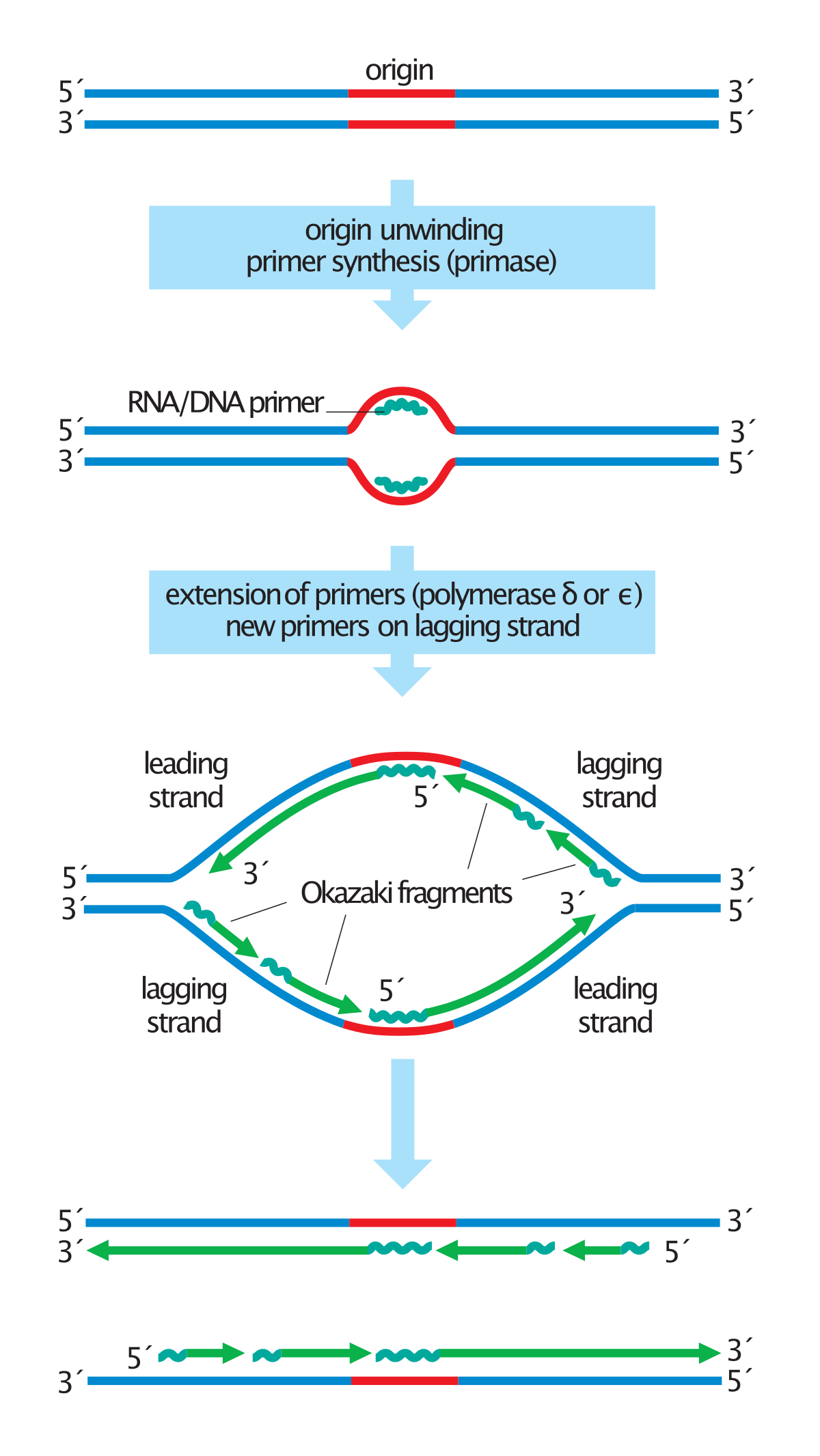
Okazaki Fragments Wikipedia
October 3, 1996 - About the BP Project History What People Say Kudos & Awards Project Team Site Credits Copyright Statement Linking to Site BP Style Manual Instructional Design · Graduate Studies in The Life Sciences
Sdjohnston Faculty Noctrl Edu
Roles of DNA polymerases and other replication enzymes. Leading and lagging strands and Okazaki fragments.
Dna Replication And Repair Dna Replication Sparknotes
A moving replication fork. (A) This schematic diagram shows a current view of the arrangement of replication proteins at a replication fork when the fork is moving. The diagram in Figure 5-21 has been altered by folding the DNA on the lagging strand to ...
Dna Replication Definition Enzymes Steps Mechanism Diagram
Replication Fork. The replication fork is a structure which is formed during the long helical DNA during the process of DNA replication. It is activated by helicases, which helps in breaking the hydrogen bonds, which holds the two strands of the helix. The resulting structure has two branching’s which is known as prongs, where each one is ...
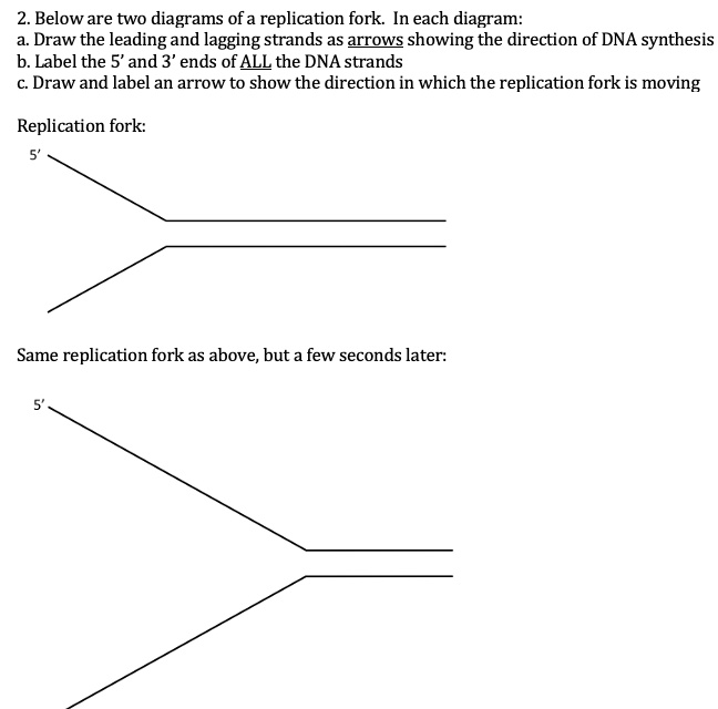
Solved 2 Below Are Two Diagrams Ofa Replication Fork In Each Diagram A Draw The Leading And Lagging Strands As Arrows Showing The Direction Of Dna Synthesis B Label The 5 And 3
1. Below is a replication fork (one side of an origin): a double-stranded DNA partially opened up to provide single-stranded regions where replication can occur. Draw and label the following: RNA primers (label 3′ and 5′ ends), leading-strand DNA polymerase and new DNA (label 3′ and 5′ ends and show direction of synthesis
Nucleic Acids And The Genetic Material Problem Set
In this diagram of the process of DNA replication at a replication fork, the strand labeled B is the: A template strand. B lagging strand. C leading strand. D Okazaki fragment. E RNA primer. The Biology Project University of Arizona Thursday, October 3, 1996 Contact the Development Team.
A Schematic Representation Of The Replication Fork Polymerase Iii Download Scientific Diagram
Our videos prepare you to succeed in your college classes. Let us help you simplify your studying. If you are having trouble with Chemistry, Organic, Physics, Calculus, or Statistics, we got your back! Our videos will help you understand concepts, solve your homework, and do great on your exams.
Dna Replication Steps Diagram Expii
Draw a labeled diagram of a replicating fork. Hint: Replicating fork is the structure of the DNA double helix after the unzipping by ligase enzyme. This leads to two strands called leading and lagging strands. DNA replication is the process of duplication of DNA during cell division. DNA is a self-replicating structure and the semi-conservative ...
Dna Replication Cell Biology Flashcards Draw It To Know It
21 Jul 2021 — One of the strands is oriented in the 3' to 5' direction (towards the replication fork), this is the leading strand?. The other strand is ...
Dna Replication
November 1, 2016 - DNA replication has been well studied in bacteria primarily because of the small size of the genome and the mutants that are available. E. coli has 4.6 ...
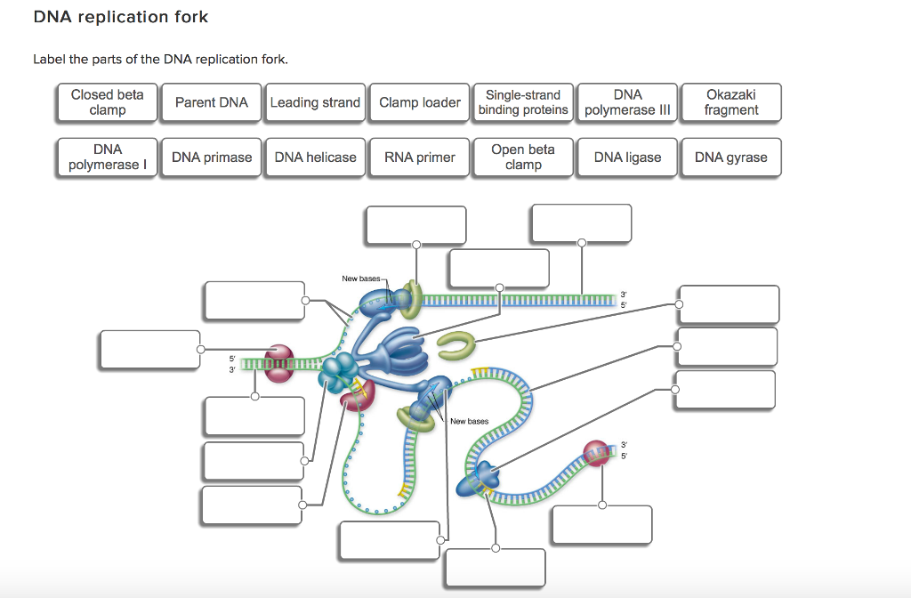
Solved Label The Parts Of The Dna Replication Fork Chegg Com
replication fork: the Y-shaped structure formed during the initiation of replication semiconservative replication: the method used to replicate DNA in which the double-stranded molecule is separated and each strand acts as a template for a new strand to be synthesized, so the resulting DNA molecules are composed of one new strand of nucleotides ...

Nucleic Acids And The Genetic Material Problem Set

Dna Replication Microbiology

File Replication Fork Svg Wikimedia Commons
Okazaki Fragments
9 2 Dna Replication Concepts Of Biology 1st Canadian Edition
Molecular Mechanism Of Dna Replication Article Khan Academy

Bioexcel 190 Molecular Genetics Key
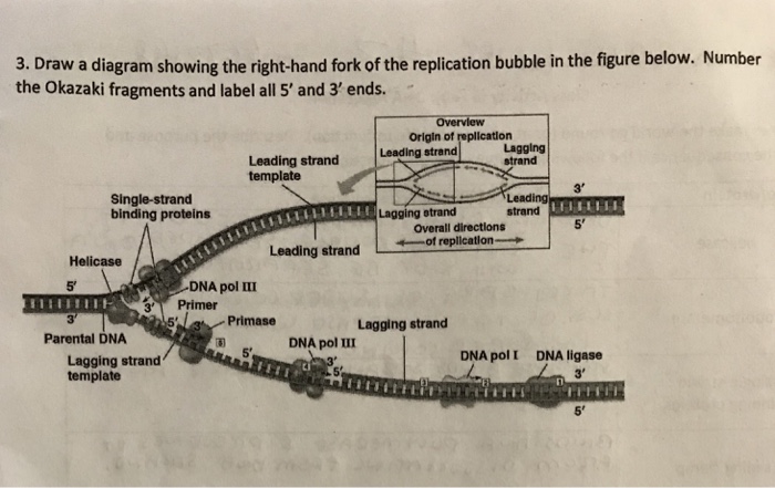
Solved Draw A Diagram Showing The Right Hand Fork Of The Chegg Com
1
1
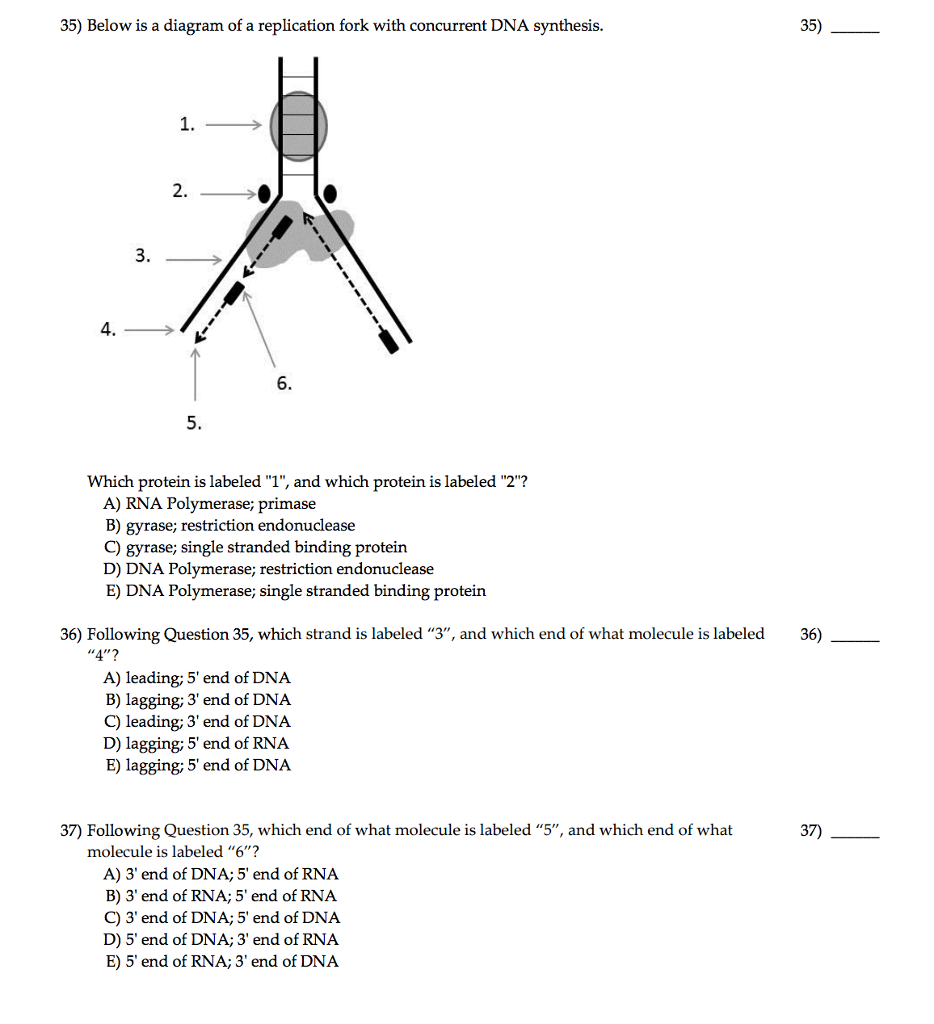
Solved Below Is A Diagram Of A Replication Fork With Chegg Com


Comments
Post a Comment