39 neuromuscular junction diagram labeled
A neuromuscular junction between a motor neuron and skeletal muscle cell. Synaptic transmission includes all the events within the synapse leading to excitation of the muscle. Let me make a quick ... First, the nerve and muscle cells must make contact, yet the two cells don't actually touch. The junction between a neuron and a muscle fiber is called the neuromuscular junction (NMJ) (see Figure 8.3). The junction, just as in the junction between neurons, is called a chemical synapse, and there is always a space between the cells called a ...
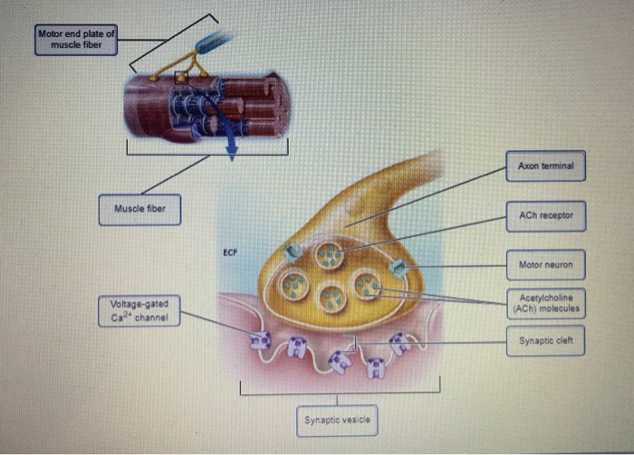
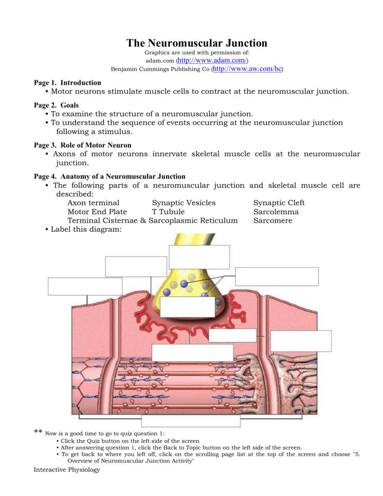
Anatomy of a Neuromuscular Junction • The following parts of a neuromuscular junction and skeletal muscle cell are described: Axon terminal Synaptic Vesicles Synaptic Cleft Motor End Plate T Tubule Sarcolemma Terminal Cisternae & Sarcoplasmic Reticulum Sarcomere • Label this diagram: ** Now is a good time to go to quiz question 1:
Neuromuscular junction diagram labeled
The neuromuscular junction (NMJ) is a synaptic connection between the terminal end of a motor nerve and a muscle (skeletal/ smooth/ cardiac). It is the site for the transmission of action potential from nerve to the muscle. It is also a site for many diseases and a site of action for many pharmacological drugs.[1][2][3][4] In this article, the NMJ of skeletal muscle will be discussed. Sep 30, 2021 · The neuromuscular junction then, is a key component in the body’s ability to produce and control movement. Amazingly, processes at the neuromuscular junction take place at speeds that allow movements to occur with no appreciable delay or lag. This article will discuss the anatomy and function of the neuromuscular junction. A neuromuscular junction (NMJ), also called a myoneural junction, is the connection between a motor neurons and a muscle fibers. These neurons are the site at which the neuron transmits a signal from the brainto the muscle fiber, causing it to contract. Therefore, neuromuscular junctions represent the channel of communication between the nervous systemand muscle cells. Their function is to allow the nervous system to control the contraction of muscles, and hence they represent an important structure in the regulation of much of our biological functions.
Neuromuscular junction diagram labeled. Draw and label a diagram of a motor unit. Terms to know: dendrite cell body (soma) nucleus axon motor end plate synapse. Neuromuscular Function Draw and label a diagram of a motor unit. AKA neuromuscular junction. Quick Muscle Structure Review. Source: Boundless. "Motor Units." Boundless Anatomy and Physiology. Boundless, 27 Sep. 2016 ... with surrounding intercellular fluid, The junction between a neuron and a muscle is called a(an) junction, is the neurotransmitter, At a neuromuscular junction, acetylcholine released from a(an) depolarizes the muscle cell membrane and triggers muscle contractions. Neuromuscular Junction Label the following parts of a neuromuscular junction, 4.1 Anatomy of the Neuromuscular Junction. The synapse for which most is known is the one formed between a spinal motor neuron and a skeletal muscle cell. Historically, it has been studied extensively because it is relatively easy to analyze. However, the basic properties of synaptic transmission at the skeletal neuromuscular junction are very ... Neuromuscular Junction Structure and Functions. The synapse or connection between a motor neuron and a skeletal muscle is known as neuromuscular junction. Communication happens between the neuron and muscle via nerve cells. Due to this communication or transmission of signal, the muscle is able to contract or relax.
3. $10.00. PDF. In this activity students will follow a procedure that instructs them to color and label 4 different sheets: (1) The neuromuscular junction (2) The sarcoplasmic reticulum (3) The sarcomere (4) The cross-bridge cycleAs students color and label each they will also address each step of the muscle contracti. Coloring Exercise 4-2 The Neuromuscular Junction COLORING INSTRUCTIONS Color each structure and its name at the same time, us-ing the same color. 1. Color the cytoplasm of the muscle cell light pink; color the nuclei purple. 2. Color the column of sar-comeres indicated by the bracket. 3. In the other sarcomeres, color the actin filaments NEUROMUSCULAR JUNCTION 1. DR NILESH KATE MBBS,MD ASSOCIATE PROF DEPT. OF PHYSIOLOGY NEURO- MUSCULAR JUNCTION. 2. OBJECTIVES. To draw the schematic diagram of Neuro-muscular junction. To describe the events of Neuromuscular transmission Classify neuromuscular blockers & give mechanism of action Name common disorders of neuromuscular j Question of previous year • Neuromuscular junction- (5m)- aug 2006 • Draw a neat diagram of neuromuscular junction. Describe the transmission of impulse across neuromuscular junction- (10m)- jan 2008
A neuromuscular junction (or myoneural junction) is a chemical synapse between a motor neuron and a muscle fiber.. It allows the motor neuron to transmit a signal to the muscle fiber, causing muscle contraction.. Muscles require innervation to function—and even just to maintain muscle tone, avoiding atrophy.In the neuromuscular system nerves from the central nervous system and the peripheral ... Click here👆to get an answer to your question ️ (a) Draw a labelled diagram of neuromuscular junction.(b) Compare nervous and hormonal mechanisms for control and co - ordination in animals. Start studying neuromuscular junction labeling. Learn vocabulary, terms, and more with flashcards, games, and other study tools. About this Quiz. This is an online quiz called Neuromuscular Junction Labeling. There is a printable worksheet available for download here so you can take the quiz with pen and paper.
Neuromuscular Junction Parts Structure And Steps Kenhub
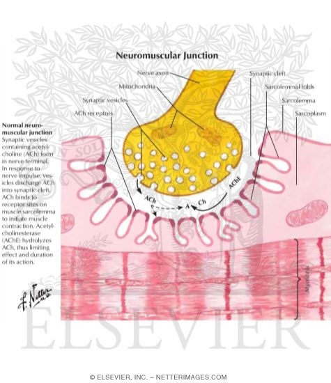
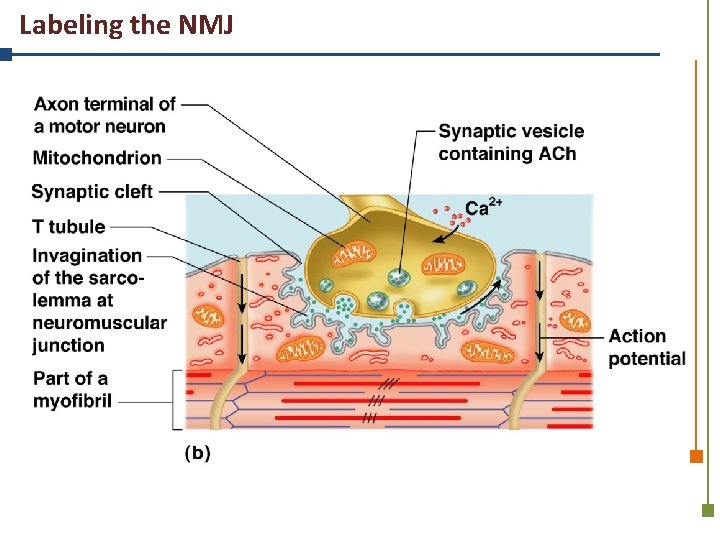
Figure 9.8 Events at the Neuromuscular Junction (2 of 4) Ca2+ Axon terminal of motor neuron Synaptic vesicle containing ACh Mitochondrion Synaptic cleft Junctional folds of sarcolemma Fusing synaptic vesicles ACh Sarcoplasm of muscle fiber Ca2+ 1 2 3 4
1
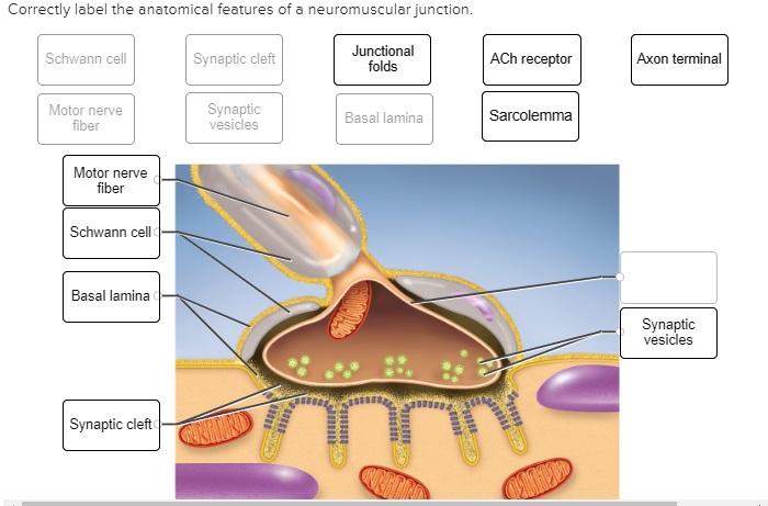
Anatomy and Physiology; Anatomy and Physiology questions and answers; Label the parts of the neuromuscular junction. Drag the labels onto the diagram to identify parts of the neuromuscular junction. Question: Label the parts of the neuromuscular junction. Drag the labels onto the diagram to identify parts of the neuromuscular junction.

Neuromuscular Junction The Neuromuscular Junction Is A Specialized Download Scientific Diagram
Muscle and Neuromuscular Junction Peter Takizawa Department of Cell Biology •Types and structure of muscle cells •Structural basis of contraction •Triggering muscle contraction. Skeletal muscle consists of bundles of long, multinucleated cells.

The Neuromuscular Junction Described Concisely Youtube
NEUROMUSCULAR JUNCTION LABELING (6 POINTS) Label the axon, motor end plate (neurotransmitter receptor), calcium channel, synaptic vesicles, neurotransmitter, and synapse in the diagram below. 1. Neurotransmitter 2. synapse 3. Motor end plate 4. Calcium channel 5. Synaptic vesicles 6. Axon Answer these questions: 1.
Solved Motor End Plate Of Muscle Fiber Axon Terminal Muscle Chegg Com
Aug 03, 1974 · Neuromuscular Junction Labeling (6 points) Label the axon, motor end plate (neurotransmitter receptor), calcium channel, synaptic vesicles, neurotransmitter, and synapse in the diagram below. 1. Neurotransmitter 2. Synapse 3. (Neurotransmitter receptor) Motor end plate 4. Calcium channel 5. Synaptic vesicles 6.

Neuromuscular Junction Labeling Diagram Quizlet
http://armandoh.org/Talks about the space between a neuron and muscle, and describes with a bit of detail about this relationship.https://www.facebook.com/Ar...
1
Nov 25, 2020 · Neuromuscular junction is a microstructure present at the junction of motor neurons and the skeletal muscle fibers. It acts as a bridge connecting the skeletal system and the nervous system. The neuromuscular junction is a chemical synapse. The presynaptic terminal is the axonal terminal of. motor neuron containing synaptic vesicles.
Solved Correctly Label The Anatomical Features Of A Chegg Com
In this video , i have explained the structure , function of neuromuscular junction and how impulse is transferred through it into the muscle . Acetylcholine...
2
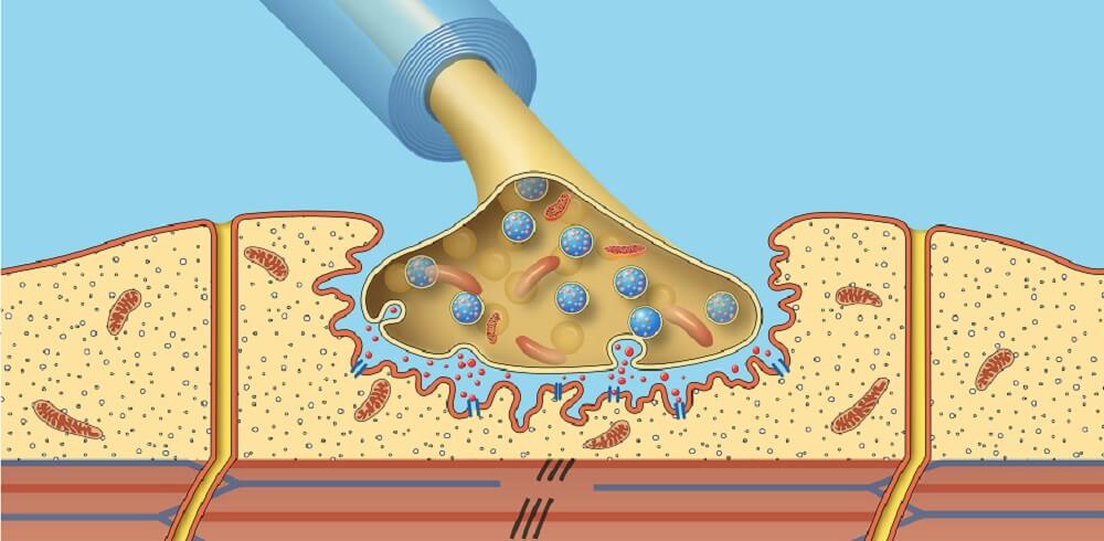
Structure of the Neuromuscular Junction. Most axons of peripheral nerves terminate on muscle cells. Whereas terminals of autonomic nerve fibers do not come in intimate contact with smooth muscle or gland cells, terminals of motor fibers form large synapses with muscle fibers, called neuromuscular junctions or motor end plates (Fig. 1).

Acetylcholine Vector Illustration Labeled Scheme With Neurotransmitter Acetylcholine Vector Illustration Labeled Scheme Canstock
Neuromuscular Junction. A neuromuscular junction (or myoneural junction) is a chemical synapse formed by the contact between a motor neuron and a muscle fiber. 3. Axon Terminal (1) The somewhat enlarged, often club-shaped endings by which axons make synaptic contacts with other nerve cells or with effector cells. Also called, presynaptic terminal.

Illustration Of A Neuromuscular Junction Download Scientific Diagram
Labeled diagram with neuromuscular junction, glandular and other neirons example. Closeup with isolated axon, cleft and dendrite structure. Endoderm, mesoderm and ectoderm vector illustration labeled infographic diagram. Isolated germ, stomach, pancreatic and lung cells. Pigment, skin and neurons of brain scheme.

Frontiers Fine Localization Of Acetylcholinesterase In The Synaptic Cleft Of The Vertebrate Neuromuscular Junction Molecular Neuroscience
A neuromuscular junction (NMJ), also called a myoneural junction, is the connection between a motor neurons and a muscle fibers. These neurons are the site at which the neuron transmits a signal from the brainto the muscle fiber, causing it to contract. Therefore, neuromuscular junctions represent the channel of communication between the nervous systemand muscle cells. Their function is to allow the nervous system to control the contraction of muscles, and hence they represent an important structure in the regulation of much of our biological functions.

Lap Proteins Are Localized At The Post Synaptic Membrane Of Neuromuscular Junctions And Appear To Modulate Synaptic Morphology And Transmission Kravic 2016 Journal Of Neurochemistry Wiley Online Library
Sep 30, 2021 · The neuromuscular junction then, is a key component in the body’s ability to produce and control movement. Amazingly, processes at the neuromuscular junction take place at speeds that allow movements to occur with no appreciable delay or lag. This article will discuss the anatomy and function of the neuromuscular junction.
Myasthenia Gravis
The neuromuscular junction (NMJ) is a synaptic connection between the terminal end of a motor nerve and a muscle (skeletal/ smooth/ cardiac). It is the site for the transmission of action potential from nerve to the muscle. It is also a site for many diseases and a site of action for many pharmacological drugs.[1][2][3][4] In this article, the NMJ of skeletal muscle will be discussed.

Acetylcholine Neurotransmission Section 1 Chapter 11 Neuroscience Online An Electronic Textbook For The Neurosciences Department Of Neurobiology And Anatomy The University Of Texas Medical School At Houston

Neuromuscular Junction Vector Illustration Scheme Labeled Cell Infographic Stock Illustration Download Image Now Istock

Junctionopathies Disorders Of The Neuromuscular Junction Veterian Key
Neuromuscular Junction The Definitive Guide Biology Dictionary
The Neuromuscular Junction

Art Labeling Activities

Neuromuscular Junctions And Muscle Contractions Anatomy And Physiology I

Clip Art Vector Neuromuscular Junction Vector Illustration Scheme Labeled Cell Infographic Stock Eps Gg107036885 Gograph
1

End Plate Potential Wikipedia

Neuromuscular Junction Diagram Quizlet

Neuromuscular Junction Vector Illustration Scheme Labeled Cell Infographic Art Print Barewalls Posters Prints Bwc60977910

The Composition Development And Regeneration Of Neuromuscular Junctions Sciencedirect
2

Draw And Label A Diagram Of A Motor Unit Ppt Video Online Download
Neuromuscular Junction The Definitive Guide Biology Dictionary
Draw A Neat And Well Labelled Diagram Of The Neuromuscular Junction Sarthaks Econnect Largest Online Education Community

Motor Neurone Labeled Royalty Free Cliparts Vectors And Stock Illustration Image 44483443
3

03 05 Neuromuscular Junction Labeling 6 Points Label The Axon Motor End Plate Neurotransmitter Receptor Calcium Channel Synaptic Vesicles Course Hero
Anatomy Physiology Park Tudor School Skeletal Muscle Activity

Label The Axon Motor End Plate Neurotransmitter Receptor Calcium Channel Synaptic Vesicles Neurotransmitter And Synapse In The Diagram Below Course Hero

Neurons Synapses And Communication Plasma Membrane 78 Steps Health

Lesson 3 05 Nerve Conduction Pdf Neuromuscular Junction Labeling 6 Points Label The Axon Motor End Plate Neurotransmitter Receptor Calcium Channel Course Hero

Medical K I S S Neuromuscular Junction


Comments
Post a Comment