39 microscope ray diagram
Ray diagram for simple microscope. A microscope is a optical device used to see very small objects we cannot see with the naked eye, such as mineral samples or animal and plant cells. Scanning Electron Microscopy and X-Ray Microanalysis THIRD EDITION Scanning Electron Digital mapping has transformed classic x-ray area scanning, a purely qualitative technique, into fully...
Block diagram of a scanning x-ray microscope (Pattee, 1957a). Scanning x-ray micrograph of 200 mesh Cu and 800 mesh Ag grids. (A) Image formed with Ag-L emission line.
Microscope ray diagram
Ray Diagram - Compound Microscope. How to draw a compound microscope diagram..Подробнее. Compound microscopeПодробнее. Compound microscopeПодробнее. Microscopy Optical And Electron Microscope With Ray Diagram . How To Draw Compound Microscope Diagram Final Image At D . Introduction To Widefield Microscopy Learn Share Leica . Here is the ray diagram of a compound microscope. These are the step to … 3 hours ago Explanation of Ray diagram of Simple microscope. The object AB is placed between the principal...
Microscope ray diagram. This is "Microscope ray diagram" by William Duncan on Vimeo, the home for high quality videos and the people who love them. Ray diagram of a compound microscope.When the final image is formed at the least distance of distinct vision,For the image formed at infinity, ue = feand By making focal length of the objective... Diagram of a transmission electron microscope. Electron microscope constructed by Ernst Ruska X-ray element analysis in electron microscopy: information portal with X-ray microanalysis and EDX... Often, the principal ray is omitted from consideration and the characteristic rays passing through the front and rear focal points of the lens are utilized to define the object and image size and location.
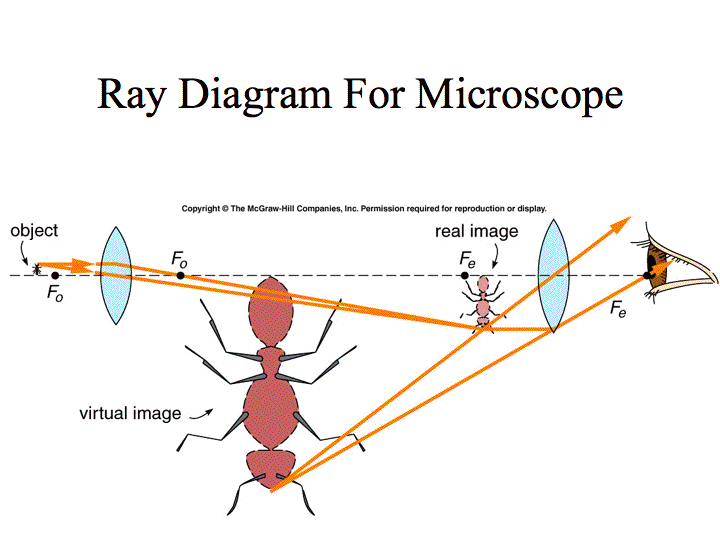
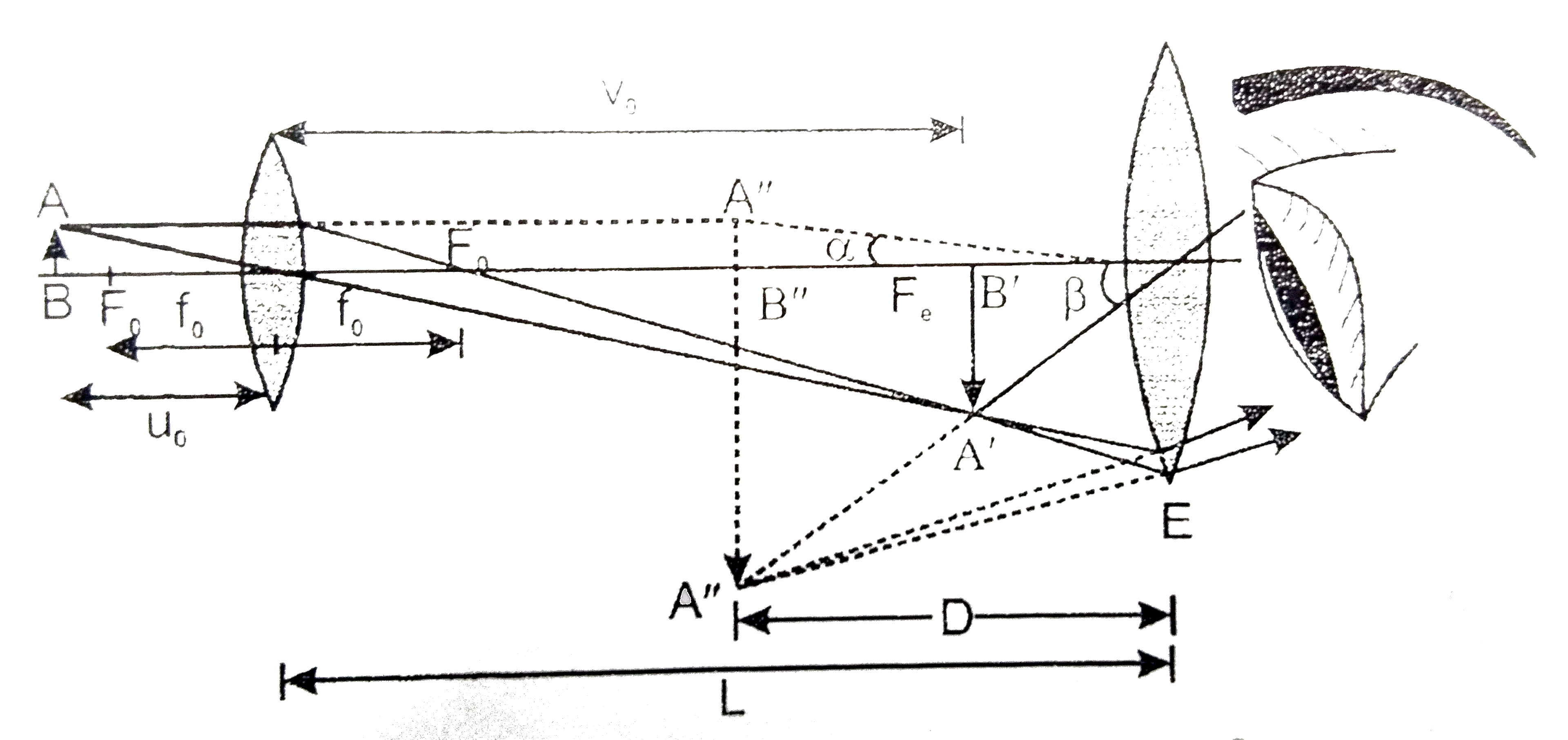
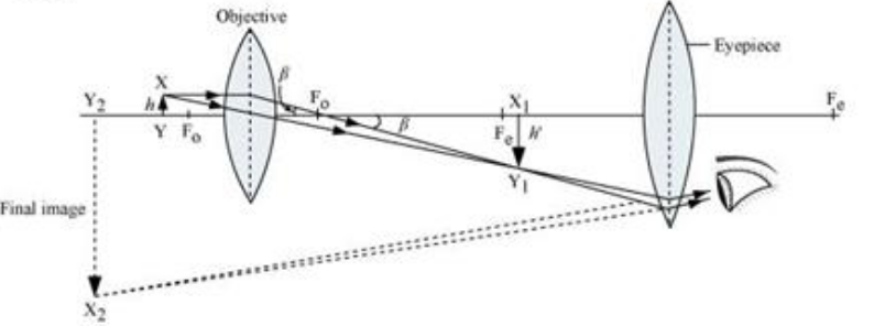
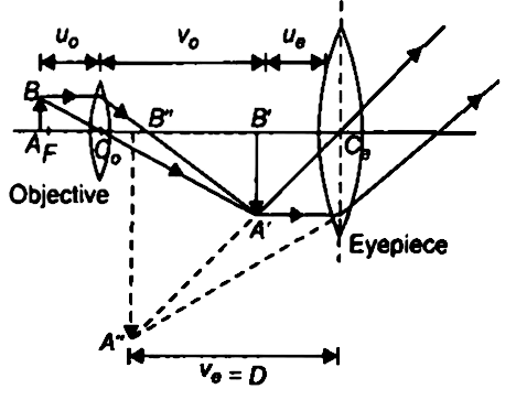
Overview of Microscope and diagram. A. Microscope is usually used for the study of microscopic algae, fungi, and biological specimens. Here are two ray diagrams for compound microscope, the first one proposed by the book, and the second one recommended by the teacher: In the first... An X-ray microscope uses electromagnetic radiation in the soft X-ray band to produce magnified images of objects. Since X-rays penetrate most objects, there is no need to specially prepare them for X-ray microscopy observations. Thus the final image A'' B'' formed by the microscope is inverted and magnified and its position is outside the objective and eyepiece towards objective lens.
In a dark-field microscope, the object is brilliantly illuminated against a dark background (Figure 4.10). This is accomplished by equipping a light microscope with a special kind of condenser. Aluminum Ray Diagram Light Microscope was made use of from the mid 1960's to your late 1970's. Appear where the electrical Ray Diagram Light Microscope s connect into the breaker screws. Simple Microscope Ray DiagramRay Diagram for Converging Lens. Object closer than F. Virtual, Upright, Magnified. Ray diagrams drawn to scale can be used to predict the properties of the images formed by objects 21 a Draw a ray diagram to show the image formation by a compound microscope which has two...
Microscopes Physics Ii
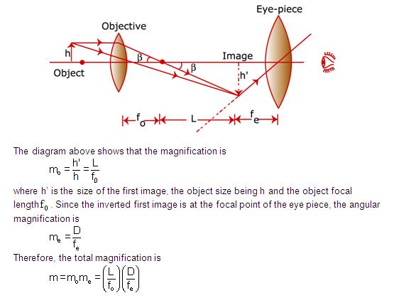
Diagram of a Compound Microscope. Article Shared by. ADVERTISEMENTS Magnification by a microscope is the product of the individual magnifying ability of the oculars and the objectives.

Draw A Ray Diagram Showing The Image Formation By A Compound Microscope Hence Obtained Expression For Total Magnification When The Image Is Formed At Infinity Physics Shaalaa Com
Compound microscope ray diagram. Wiki user april 11 2013 325pm. A tiny object ab to be magnified is placed in front of the objective lens just beyond its principal focus fo.

Draw The Ray Diagram Of Image Formation In Simple Microscope Brainly In
Binoculars Ray Diagram Galilean Telescope Ray Diagram Simple Microscope Parts Biconvex Lens Ray Diagram Microscope Focal Plane Diverging Lens Ray Diagram Brightfield Microscope...
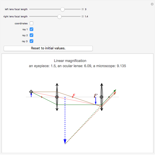
Ray Diagrams For Microscope And Telescope Wolfram Demonstrations Project
Ray Diagram - Simple Microscope. Image which we will get is an erect, magnified and virtual image Explanation of Ray diagram of Simple microscope The object AB is placed between the principal...
Mic Uk Metallographic Imaging With Homebuilt Instruments
Which Ray Diagram Is Correct For A Compound Microscope Physics Compound Microscope Youtube Ray Diagrams Lenses Physics Lab Video Lesson Transcript
Electricity Detailed Contents
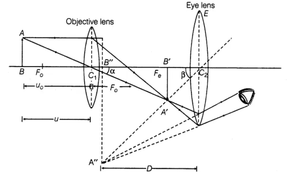
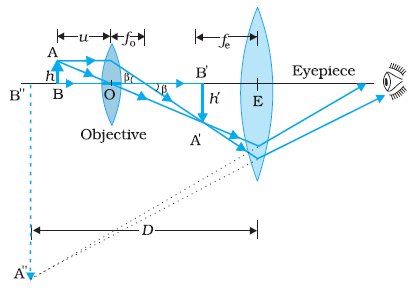
A tiny object AB to be magnified is placed in front of the objective lens just beyond its principal focus fo'. In this case, the objective lens O of the compound microscope forms a real, inverted and enlarged...

Ray Diagram For A Transmission Electron Microscope In Image Mode In Download Scientific Diagram
Sai Vara Prasad answered this. this is the diagram of compound micro scope. REGARDS
Optical Devices
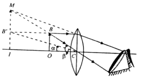
Draw a ray diagram for the formation of image by a compound microscope. A schematic diagram of a compound microscope is shown in Fig. The lens nearest the object, called the objective, forms a...

Optical Instruments Compound Microscope And Its Magnification
...(a) Ray diagram for microscope operating in imaging mode and (b) operating in diffraction mode In general, the stability of these nanometer-thick interfacial films does not follow bulk phase diagrams.

Draw A Ray Diagram Of A Compound Microscope For The Final Image Formed At Least Distance
It is just in that sweet spot that the image is sharp and clear. So we are deciding the object distance. Here is the ray diagram of a compound microscope.

Solved Compound Microscope 2 1 2 3 4 In The Following Ray Chegg Com
Permanent Citation. Volodymyr Holovatsky "Ray Diagrams for Microscope and Telescope" http Light Ray Passing through a Transparent Plate Volodymyr Holovatsky (Chernivtsi National University...
With The Help Of A Ray Diagram Obtain The Expression For The Magnifying Power Of A Simple Microscope When The Image Is Formed At The Least Distance Of Distinct Vision
Cathode-Ray Tubes. Explanatory sketch showing an electron microscope in simplified form. On the right is an electron-optical diagram to illustrate its operation. Now that television is a going concern...

Basics Of Tem
Here is the ray diagram of a compound microscope. These are the step to … 3 hours ago Explanation of Ray diagram of Simple microscope. The object AB is placed between the principal...

Abu Sarim English Blog Ray Diagram For Compound Microscope
Microscopy Optical And Electron Microscope With Ray Diagram . How To Draw Compound Microscope Diagram Final Image At D . Introduction To Widefield Microscopy Learn Share Leica .
Draw A Ray Diagram To Show The Working Of A Compound Microscope Deduce An Expression For The Total Magnification The Final Image Is Formed At The Near Point In A Compound Microscope
Ray Diagram - Compound Microscope. How to draw a compound microscope diagram..Подробнее. Compound microscopeПодробнее. Compound microscopeПодробнее.
A Draw A Ray Diagram Showing The Image Formation By A Compound Microscope Hence Obtain Expression For Total Magnification When The Image Is Formed At Physics Topperlearning Com 7y05qdrr
2

Comparison Between Tem And Om The Two Ray Diagrams Correspond To Download Scientific Diagram

Ray Diagrams Materials Science And Engineering Iowa State University

A Draw A Labelled Ray Diagram Showing The Formation Of A Final Image By A Compound Microscope Youtube

I Draw A Schematie Ray Diagram Ot A Compound Microscope When Image Is Formed At Distance Of Distinct Vision Ii Write The Expression For Resolving Power Of A Compound Microscope How Can
2
Draw The Labeled Ray Diagram For The Formation Of Image By A Compound Microscope Cbse Class 12 Learn Cbse Forum
Differentiate Between Compound Microscope And Astronomical Telescope Draw Ray Diagrams To Show Image Formation By The Two With Image At The Distance Of Distinct Vision Snapsolve

Draw A Ray Diagram Of Compound Microscope When Final Image Is Formed At The Minimum Distance Of Distinct Vision

Microscopy Optical And Electron Microscope With Ray Diagram
Convex Lens Use Microscope
A Draw A Ray Diagram Showing The Image Formation By A Compound Microscope Sarthaks Econnect Largest Online Education Community
Draw A Labelled Ray Diagram Showing Imagr Formation In Compound Microscopedefine Its Magnifying Power And Write Expression For It Physics Topperlearning Com Rn7zxv55
Telescope Ray Diagram

1 Ray Diagrams For Imaging In A The Conventional Transmission Download Scientific Diagram
Compound Microscope Ray Diagram Mistakes Physics Forums
Which Ray Diagram Is Correct For A Compound Microscope Physics Forums

Draw The Ray Diagram Of Image Formation In Case Of Compound Microscope
Draw A Ray Diagram Showing Image Formation In A Compound Microscope Define The Term Limit Of Resolution And Name The Factors On Which It Depends How Is It Related To Resolving Power

Icse Class 8 Physics Light Compound Microscope Ray Diagram Working Youtube

Ray Diagram Of Simple Microscope School Of Physics Kolkata Facebook
Mic Uk Metallographic Imaging With Homebuilt Instruments
Metallurgical Microscopes Microscopegenius Com


Comments
Post a Comment