38 drag the labels onto the diagram to identify the various types of synarthroses and amphiarthroses.
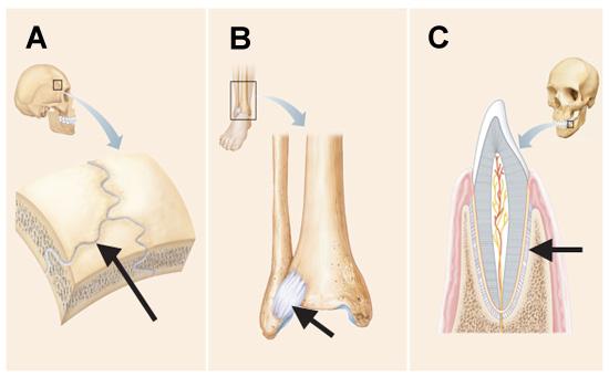
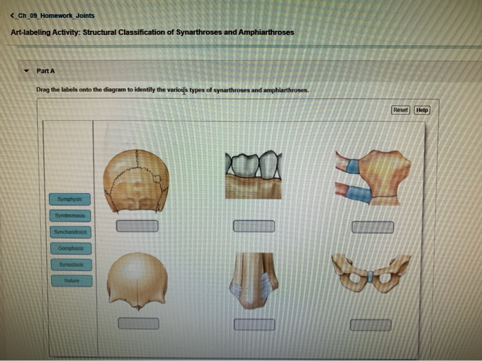
4 Examining the Histologic Structure of the Large Intestine 584 5 Identifying Types of Teeth 585 6 Studying Microscopic Tooth Anatomy 585 7 Examining Salivary Gland Tissue 586 8 Examining the Histology of the Liver 587 Review Sheet 589 39 Digestive System Processes: Chemical E x ercise and Physical 595 The functional classification divides joints into three categories: synarthroses, amphiarthroses, and diarthroses: Synarthroses are a joints that are immovable. This includes sutures, gomphoses, and synchondroses. Amphiarthroses are joints that allow slight movement, including syndesmoses and symphyses. Diarthroses are joints that allow for ...
Science. Anatomy and Physiology. Anatomy and Physiology questions and answers. Part A Drag the labels onto the diagram to identify the various types of synarthroses and amphiarthroses. Reset Help Syndesmosis Symphysis Suture Synostosis Gomphosis Synchondrosis.

Drag the labels onto the diagram to identify the various types of synarthroses and amphiarthroses.
HW 4 Due: 11:59pm on Friday, October 6, 2017 To understand how points are awarded, read the Grading Policy for this assignment. Art-labeling Activity: Curves and Regions of the Vertebral Column Learning Goal: To learn the curves and regions of the vertebral column. Label the curves and regions of the vertebral column. Part A Drag the labels onto the diagram to identify the curves and regions ... Drag the labels onto the diagram to identify the various types of synarthroses and amphiarthroses. 1. Suture 2. Gomphosis 3. Synchodrosis 4. Synostosis hyaline cartilage. Which component of the connective tissue in this field of view is highlighted? extracellular matrix. The type and characteristics of a given joint determines its degree and type of movement. Joints can be classified based on structure and function. Structural classification of joints categorizes them based on the type of tissue involved in formation. There are three structural classifications of joints: fibrous, cartilaginous, and synovial.
Drag the labels onto the diagram to identify the various types of synarthroses and amphiarthroses.. diarthrosis: a joint that can move freely in various planes; synarthrosis: immovable joint in which two bones are connected rigidly by fibrous tissue; amphiarthrosis: slightly movable joint in which the surfaces of bones are connected by ligaments or cartilage Label each of the following systems as high or active site and changes the conformation low entropy: i. the instant that a perfume bottle is of the active site, decreasing its affinity sprayed compared with 30 seconds later, ii. an old for the substrate 1950s car compared with a brand new car, and iii. d. Binds directly to the active site and a ... Identify and describe the different types of mechanical loads that act on the human body. Identify and describe the uses of available instrumentation for measuring kinetic quantities. Distinguish between vector and scalar quantities. Solve quantitative problems involving vector quantities using both graphic and trigonometric procedures. Terry R. Martin_ Cynthia Prentice-Craver - Laboratory manual for human anatomy & physiology _ main version (2019).pdf - Free ebook download as PDF File (.pdf), Text File (.txt) or read book online for free. Labaratory Manual For A&P
Nov 19, 2021 · Drag the labels onto the diagram to identify the various types of synarthroses and. These consist of the arm, located between the shoulder and elbow joints; Reasons to perform the shoulder capsular and muscular structures of the shoulder girdle. By the end of this section, you will be able to identify the following muscles. 20 Drag The Labels Onto The Diagram To Identify The . ... Label the various types of synarthroses and amphiarthroses. ... Identify the features of a typical synovial joint listed in figure 10.3 on the model, using table 10.5 and the textbook as guides; Then label them in figure 10.3. Synovial joints are joints that have a space between the adjoining bones. The functional classification divides joints into three categories: synarthroses, amphiarthroses, and diarthroses. The movement of synovial joints can be classified as one of four different types: gliding, angular, rotational, or special movement. The B cells produce specialised glycoproteins called which have 3 main functions : of particulate matter, of bacteria to facilitate their subsequent phagocytosis of cells and of toxins is the most abundant antibody. (b) Label the given diagram. ®ir Xsfc(-ai i (b) D e n a bank (d) Vijaya bank. I2>|
Academia.edu is a platform for academics to share research papers. NEW TO THIS EDITION Annotated Instructor's Edition is available as a PDF on the password-protected Instructor's Online Learning Center at www.mhhe.com/medterm3e ... A new head-to-head comparison of screening questionnaires for posttraumatic stress disorder (PTSD) shows a worrying discordance between the previous version of the PTSD definition in the ... It involves the examination of urine for the presence of normal or abnormal amounts of various elements. Substances in the urine are a prime factor in the diagnosis of diseases of the urinary system as well as of other body systems. In addition, various imaging and blood tests help diagnose conditions or diseases. 11/7/08 9:58:24 AM D.
Synovial Joints Anatomy And Physiology I
Label the various types of synarthroses and amphiarthroses. Part A Drag the labels onto the diagram to identify the various types of synarthroses and amphiarthroses. ANSWER: Correct Art-labeling Activity: The Structure of a Synovial Joint (sagittal section) Identify the structures within a synovial joint. Part A Drag the labels to identify the ...
1
Correct Part D - Summary: Components of Skin Layers Each layer of the skin is composed of a different type of tissue and contains different components. Drag and drop the labels to their appropriate location in the skin. Hint 1. Exocrine Glands of the Skin Exocrine glands secrete their products via ducts.

210 Medical Terminology Language For Healthcare Nina Thierer 0073374725 Mcgraw Hill 2010 786 9
Ashland Community & Technical College is one of the 16 colleges working to bring better lives to all Kentuckians as a part of KCTCS. ACTC currently has three campuses in Boyd County, KY that offer both on-campus and online classes. Never underestimate you.

34 Label Synovial Joint Labels Design Ideas 2020
Human anatomy [Eighth edition] 9780134243818, 1292156791, 9781292156798, 0134243811, 9780134283395, 0134283392, 9780134398303, 0134398300. For one-semester courses in human anatomy.Helps readers visualize human anatomyThe #1 best-selling book for the human an

Chapter 4 A P Lab Flashcards Quizlet
PLAY. Drag the labels onto the diagram to identify the muscle types based on fascicle organization. Drag the labels onto the diagram to identify the major skeletal muscles, anterior view. Nice work! You just studied 21 terms! Now up your study game with Learn mode.
Print A P Chapter 8 Joints Flashcards Easy Notecards
Nov 18, 2021 · Drag the labels onto the diagram to identify the various types of synarthroses and. A p2 lab 7 hw a p2 lab 6 hw a p 2 lab 5 hw a p2 lab 7 pp . Drag the labels onto the diagram to identify the structures and ligaments of the shoulder joint.

16 Anatomy Ideas Anatomy Anatomy And Physiology Medical Knowledge
Drag the labels onto the diagram to identify the various types of synarthroses and amphiarthroses. Click card to see definition 👆. Tap card to see definition 👆. 1. Suture. 2. Gomphosis. 3. Synchodrosis.

Unit 2 Exam Ch 6 7 8 Flashcards Quizlet
biochemistry and genetics contributors contributed to this project include: gi, metabolism, nutrition, endocrine, reproduction, male and female.
Drag The Labels To Identify The
The type and characteristics of a given joint determines its degree and type of movement. Joints can be classified based on structure and function. Structural classification of joints categorizes them based on the type of tissue involved in formation. There are three structural classifications of joints: fibrous, cartilaginous, and synovial.

Solved Activity 13 6 Identifying Joints Based On Their Chegg Com
Drag the labels onto the diagram to identify the various types of synarthroses and amphiarthroses. 1. Suture 2. Gomphosis 3. Synchodrosis 4. Synostosis hyaline cartilage. Which component of the connective tissue in this field of view is highlighted? extracellular matrix.

Drag The Labels Onto The Diagram To Identify The Structures And Ligaments Of The Shoulder Joint Drag The Labels Onto The Diagram To Identify The Budidayateh
HW 4 Due: 11:59pm on Friday, October 6, 2017 To understand how points are awarded, read the Grading Policy for this assignment. Art-labeling Activity: Curves and Regions of the Vertebral Column Learning Goal: To learn the curves and regions of the vertebral column. Label the curves and regions of the vertebral column. Part A Drag the labels onto the diagram to identify the curves and regions ...
Homework 1 Chapters 1 2 4 5 6 And 9 Apk 2100c Appl Human Studocu

Human Anatomy And Physiology I Post Lab 5 Homework Flashcards Quizlet

Drag The Labels Onto The Diagram To Identify The Parts And Ligaments Of The Hip Joint Homeworklib

Drag The Labels Onto The Diagram To Identify The Parts Of The Pelvis Of The Adult Homeworklib

Lab 5 Exercises 10 And 11 Flashcards Quizlet

Lab 5 Exercises 10 And 11 Flashcards Quizlet

Ap1 Lab 9 10 Hw Flashcards Quizlet

Human Anatomy And Physiology I Post Lab 5 Homework Flashcards Quizlet

Anatomy Lab 10 11 Flashcards Quizlet

Drag The Labels Onto The Diagram To Identify The Parts And Ligaments Of The Hip Joint Homeworklib

Identify The Type Of Structural And Functional Joints Chegg Com

Figure 11 1 Structural Classification Of Joints A Anterior View And B Tooth In Alveolus Diagram Quizlet

The Skeletal System Chapter Ppt Video Online Download

Articulation Joints Flashcards Quizlet
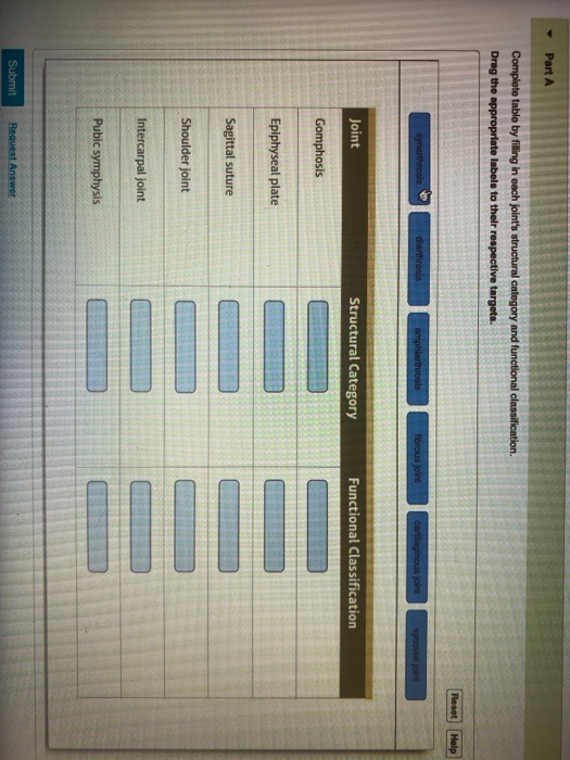
Solved Part A Complete Table By Filling In Each Joint S Chegg Com
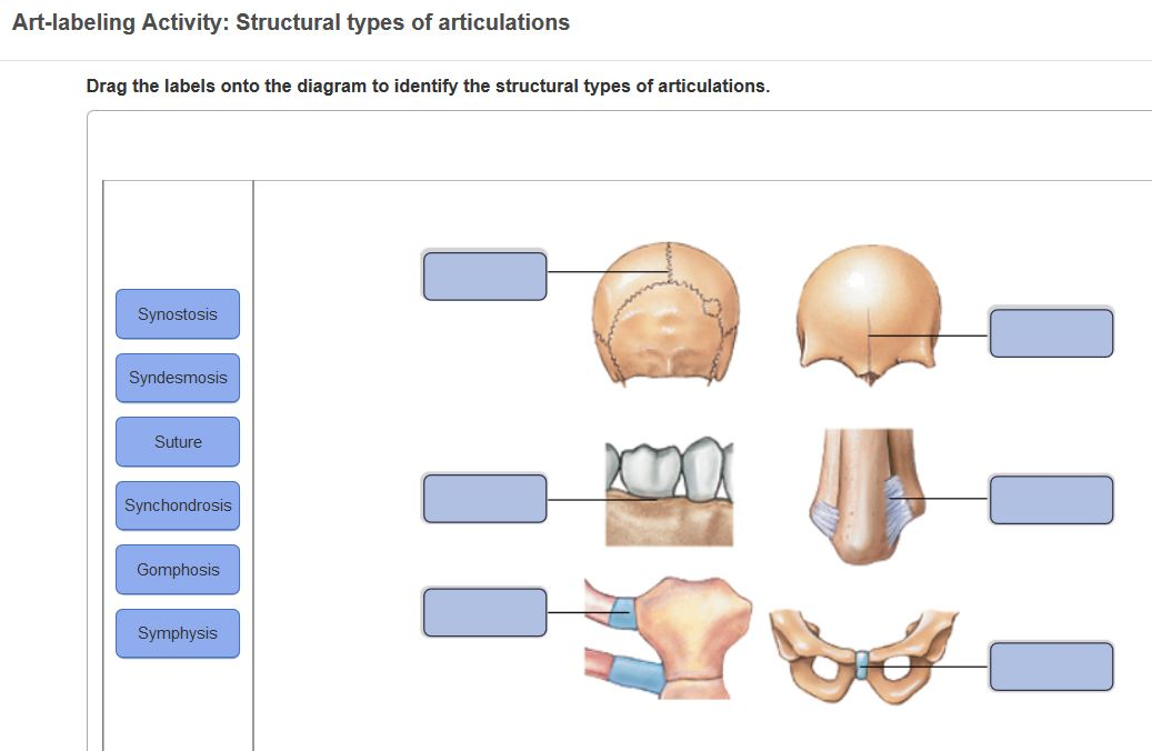
Solved Art Labeling Activity Structural Types Of Chegg Com
Napavalley Edu

Ap1 Lab 9 10 Hw Flashcards Quizlet

Anatomy Lab 10 11 Flashcards Quizlet

Chapter 9 Joint Lab Pdf 1 Anatomy I Joint Articulation Lab Names Section One Identification Which Letter Refers To The Following A Types Of Joints Course Hero
Drag The Labels Onto The Diagram To Identify The Structures And Ligaments Of The Shoulder Joint A P Exam 2 At Simmons College Studyblue Drag The Labels Onto The Diagram

Drag The Labels Onto The Diagram To Identify The Structures And Ligaments Of The Shoulder Joint Physical Therapy In Perrysburg For Shoulder
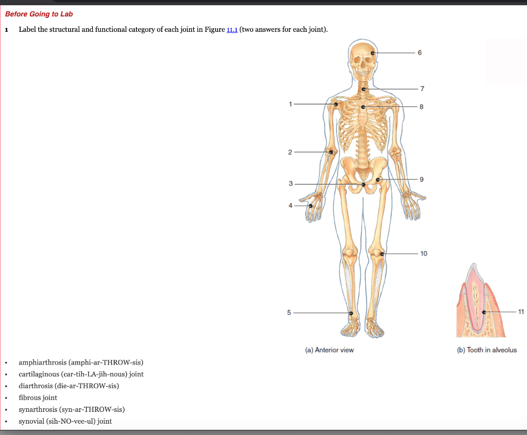
Solved Before Going To Lab 1 Label The Structural And Chegg Com

Ap1 Lab 9 10 Hw Flashcards Quizlet

Ap1 Lab 9 10 Hw Flashcards Quizlet
Solved Ch 09 Homework Joints Art Labeling Activity Chegg Com

Ap1 Lab 9 10 Hw Flashcards Quizlet


Comments
Post a Comment