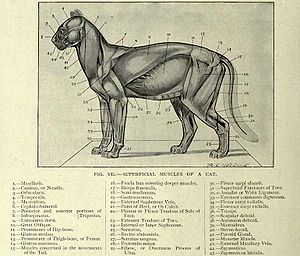
38 cat muscles labeled diagram
The male pelvis is different from a female's. An atlas of cat anatomy. #bone pelvis anatomy video #pelvic bone anatomy cat #pelvic bone anatomy in horse #pelvic bone muscle anatomy #pelvic bone teach me anatomy related posts of pelvic bone in anatomy bones in the neck This diagram of a feline skeleton shows you where all of your cat's bones ... 10,477 human skeleton labelled stock photos, vectors, and illustrations are available royalty-free. See human skeleton labelled stock video clips. of 105. skeletal system anatomy labelled skeleton skeleton label skeletal system posterior view labeled skeleton skeleton labeled skeleton labels male human skeletle system human skeletal system ...
Knee diagram tendons was posted in may 29, 2015 at 4:57 pm. Downloads diagram diagram diagram of the heart diagram definition diagramming sentences the cat 5 wiring tendon diagram will likely be your starting point to creating and setting your 1st network.

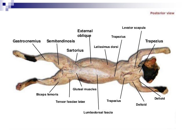
Cat muscles labeled diagram
This article lists a series of labeled imaging anatomy cases by system and modality. Brain CT head: non-contrast axial CT head: non-contrast coronal CT head: non-contrast sagittal CT head: angiogram axial CT head: angiogram coronal CT head... 11,163 spinal nerves stock photos, vectors, and illustrations are available royalty-free. See spinal nerves stock video clips. of 112. damage of nerves the spinal cord nerve and organ anatomy thorac nervous system damage dilate pupils neck nerves stenosis structure of the central nervous system spine organs. Try these curated collections. Related Posts of "Muscle Anatomy Upper Limb" Abdominal Muscle Diagram. Abdominal Muscle Diagram 12 photos of the "Abdominal Muscle Diagram" abdominal muscle anatomy bodybuilding, abdominal muscle diagram female, abdominal muscle groups diagram, human abdominal muscle diagram, lower abdominal muscle diagram, Human Muscles, abdominal muscle anatomy bodybuilding, abdominal muscle diagram female ...
Cat muscles labeled diagram. Cat Anatomy Leg Muscles from o.quizlet.com Anatomy colour diagram lasalle leg muscles sakart. Free online quiz back of leg muscle diagram. Labeled anatomy chart male back muscles stock illustration 1423699424 : Leg muscles diagram labeled : The following diagram illustrates the actions of the terms adduction, abduction, flexion and anterior ... Cross-sectional labeled anatomy of the head and neck of the domestic cat on CT imaging (bones of the skull, cervical spine, mandible, hyoid bone, muscles of the neck, nasal cavity and paranasal sinuses, oral cavity, larynx) Human body muscle diagrams. Muscle diagrams are a great way to get an overview of all of the muscles within a body region. Studying these is an ideal first step before moving onto the more advanced practices of muscle labeling and quizzes. If you're looking for a speedy way to learn muscle anatomy, look no further than our anatomy crash courses . Cow anatomy labeled diagram. Here I would like to summarize the whole anatomical features of a cow (both internal and external) with the labeled diagram. I hope you will enjoy it and learn the anatomical features of the different organs of a cow. If you need more cow-labeled diagrams, you may join with anatomy learners on social media.
Muscles Labeled Front And Back - Cat Muscles of the Lateral Surface of the Front Leg. 2.4 neck · 3 torso. Called involuntary muscles — are usually in sheets, or layers, with one layer of muscle behind the other. Leg muscle anatomical structure, labeled front, side and back view diagrams. 1.4 nose · 2 neck. Science Experiments With Cats / Schrodinger S Cat Wikipedia : Muscle Diagram To Label - Human Muscle System Functions Diagram Facts Britannica : Boy Crying Drawing Realistic / A Boy Crying : 2021 (65) Design by - Blogger Templates Class room lectures - starts from Chapter 2 in the book (here Lesson 1) Book used: Anatomy&Physiology: The Unity of Form and Function by Kenneth Saladin 6th Ed (McGrawHill) Use head phone for best audio-bear with any shake in video or listen to audio alone. Best wishes. Dr. Chandra Related Posts of "Neck Muscle Anatomy Mri" Anatomy Muscle System. Anatomy Muscle System 12 photos of the "Anatomy Muscle System" anatomy and physiology muscular system exam, anatomy and physiology muscular system labeling quiz, anatomy and physiology muscular system pdf, anatomy and physiology muscular system review, human anatomy muscular system quizzes, Human Muscles, anatomy and physiology ...
Anatomy of the dog - Illustrated atlas. This modules of vet-Anatomy provides a basic foundation in animal anatomy for students of veterinary medicine. This veterinary anatomical atlas includes selected labeling structures to help student to understand and discover animal anatomy (skeleton, bones, muscles, joints, viscera, respiratory system ... Cladogram Definition. A cladogram is the graphical representation of the hypothetical relationship (phylogenetic relationship) between different groups of organisms. It is used in the phylogenetic analysis of organisms to determine the evolutionary relationship between them. The cladogram is derived from Greek words clados and gramma where 'clados' means branch and 'gramma' means ... Anatomy Study Resources. This guide contains a variety of resources to help with the study of veterinary anatomy. Included are web resources, books (both print and electronic), and specialty resources such as flashcards. Items from the Course Reserves List for VBMS601 Gross Anatomy-Structural Adaptations to Function are marked. The cat skeletal anatomy labeled diagrams that provide in this article might help you a lot. But if you need more cat anatomy labeled diagrams, please let me known. Categories Dog and Cat Anatomy Tags cat skeleton , cat skeleton anatomy , cat skeleton anatomy labeled diagram , cat skeleton diagram , cat skeleton head , cat skeleton labeled ...
Superficial Cat Muscles Quiz
Muscle anatomy reference charts Author: Molly Smith DipCNM, mBANT • Reviewer: Dimitrios Mytilinaios MD, PhD Last reviewed: November 03, 2021 Reading time: 3 minutes If you've ever attempted to learn the origins, insertions, innervations, and functions of all 600+ muscles in the body… you'll know what a soul-destroying task it can be.

Kennedy Anne Anatomy Review
Anatomy of the cat's or dog's eye. Vertical section of the eye and eyelids. Third eyelid and Tapetum lucidum. Schematic diagram. detailed illustration. Pupil - The black opening at the centre of the iris that controls how much light is let into the eye. In bright conditions, the pupil is a small slit (see image above), in dark conditions ...

Cat Dissection Digestive Labs Ppt Video Online Download
The axial skeleton protects the brain, spinal cord, heart, lungs, and kidneys. It provides structural support and provides articulation sites to the appendicular skeleton. The axial and appendicular skeleton together make a complete human skeleton. The axial skeleton is a combination of 80 bones.

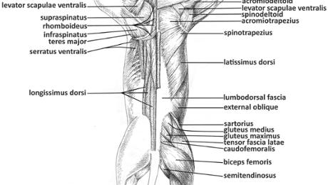
Cat Dissection Lab Muscles Of The Back And Shoulder Flashcards Quizlet
Anatomy of the Heart. Now that we have reviewed the main anatomical structures of the heart using the cartoon image, let's go back to that original diagram. You should now be able to label all the main anatomical features. Below is a blank diagram, followed by the labeled diagram with the answers.
Cat Anatomy Wikipedia
Cats and dogs do walk on their toes, in what is called digitigrade stance. This is opposed to the plantigrade stance of humans. This is shown nicely on this Wikipedia diagram, where the dog (or cat) is the middle picture, human to the left, and ungulate (hoofed animal) to the right: . The cat stands on their toe bones (phalanges).
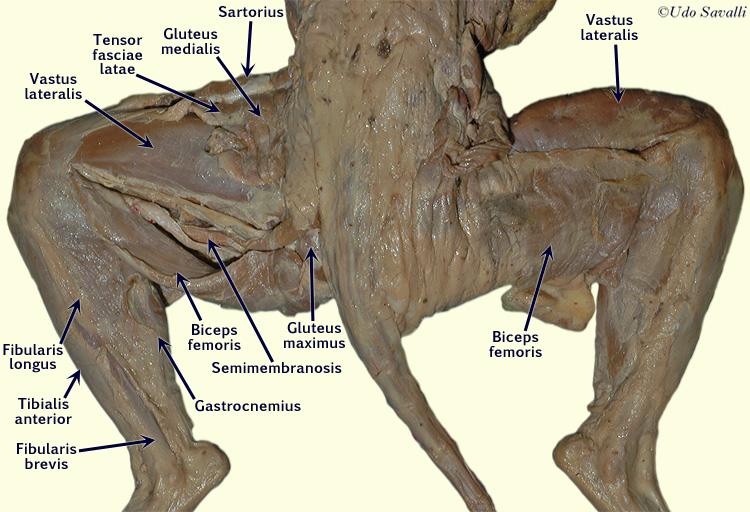
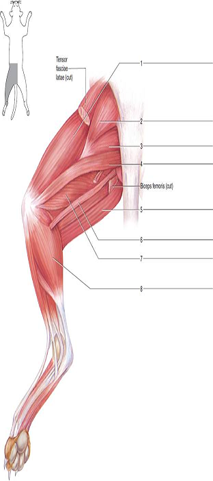
Cat Muscles Of The Lower Appendages David Fankhauser
The smooth muscles that line organs, as well as the cardiac muscle of the heart. Voluntary muscles include skeletal muscles and total about 650 in the whole human body. Muscle action can be broken up into either voluntary or involuntary action. Smooth muscle cells are tapered at the ends and do .

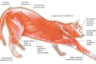
Cat Muscles
11 Cat Sleeping Positions & What They Mean . Just like their owners, cats sleep in a variety of positions. From sleeping on their side to sleeping belly up, we can learn a lot about our feline friends through their sleeping positions and habits. Many cat sleeping positions are instinctual poses from their wild ancestors and are a form of ...
Cat Muscles Of The Back Color
Cat Claws - Anatomy, Function and Disorders. The claw is a scythe-shaped appendage that is attached to the end bone of the toe. Cats have four toes on each hindfoot and five toes on the front feet. The fifth toe is the dewclaw, which is located on the inner side of the foot and does not make contact with the ground.

Anatomy Labeled Stock Illustrations 947 Anatomy Labeled Stock Illustrations Vectors Clipart Dreamstime
Cat Wallpaper Iphone - White Cat Iphone Wallpapers... 2Nd Grade Reading Lesson : Grade 2 Reading Workshe... Digestive System Circulatory System And Respirator... Labeled Cow Circulatory System Diagram / Hilum Ana... Ear Anatomy Unlabeled / Ear Structure Stock Illust... Individual Skeletal Muscle Cells : Skeletal Muscle...
Cat
Anatomy Of Pelvic Muscles Female - Pelvis And Perineum Anatomy Vessels Nerves Kenhub -. The bony pelvis is the rigid foundation to which all of the pelvic ligaments and muscles are anchored. The pelvic floor muscles also help you to control bladder and bowel function, such as allowing you to 'hold on' until an appropriate time and place.

26 Cat Muscles Ideas Cat Anatomy Muscle Anatomy Anatomy
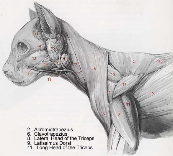
Pectoralis profundus Pectoralis minor Dog And Feline. • Origin: Most of the sternum (except its front tip) and from the surface of the front end of the abdomen (in the region of the xiphoid process). • Insertion: Upper inner surface of the humerus, and onto a vertical line on the upper third to upper half of the front of the humerus.
Bio201 Cat Muscles
Related Posts of "Muscle Anatomy Upper Limb" Abdominal Muscle Diagram. Abdominal Muscle Diagram 12 photos of the "Abdominal Muscle Diagram" abdominal muscle anatomy bodybuilding, abdominal muscle diagram female, abdominal muscle groups diagram, human abdominal muscle diagram, lower abdominal muscle diagram, Human Muscles, abdominal muscle anatomy bodybuilding, abdominal muscle diagram female ...

Cat Muscles Muscle Vet Tech Cats
11,163 spinal nerves stock photos, vectors, and illustrations are available royalty-free. See spinal nerves stock video clips. of 112. damage of nerves the spinal cord nerve and organ anatomy thorac nervous system damage dilate pupils neck nerves stenosis structure of the central nervous system spine organs. Try these curated collections.

Pin On Vet School
This article lists a series of labeled imaging anatomy cases by system and modality. Brain CT head: non-contrast axial CT head: non-contrast coronal CT head: non-contrast sagittal CT head: angiogram axial CT head: angiogram coronal CT head...
2

Cat Muscle Dissection Muscles Of Chest Neck Arms And Abdomen Bulb

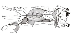
Cat Musculature Atlas Of Comparative Vertebrate Anatomy

Muscles Of The Cat Simplebooklet Com

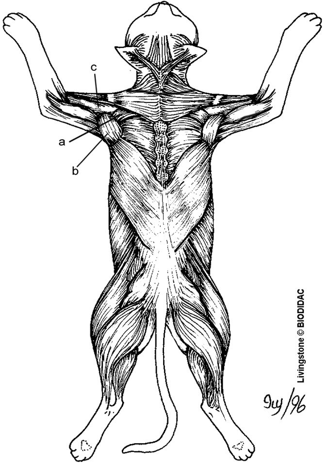
Dorsal Cat Muscles Diagram Quizlet

Cat Reference Images

Cat Muscles
2
Medical Society Takes On 2018 With Cat Dissection The Roundup

On The Cutting Edge Ap Biology Mammalian Structure And Function Dissection Kit Carolina Com
Cat Muscles Diagram Biological Science Picture Directory Pulpbits Net

Muscles Of The Cat Simplebooklet Com
Cat Muscles Lab Guide

Cat Muscles My Copy Flashcards Chegg Com
Cat Muscle Dissection Dorsal Muscles Of Back Arm Shoulder Bulb
Cat Dissection Lab Labeled Images

Cat Muscles Lab Guide

Labeled Atlas Of Anatomy Illustrations Of The Dog

Cat Muscles Ventral Superficial Shoulder Diagram Quizlet

Human Anatomy Physiology 1
Solved Identify The Numbered Muscles Of The Cat In Figures 62 24 Chegg Com
Anatomy And Physiology Lab I On Openalg

Cat Muscles Neck Diagram Quizlet

Cat Musculature Atlas Of Comparative Vertebrate Anatomy


Comments
Post a Comment